. 2020 Jul 2;10(1):10912.
doi: 10.1038/s41598-020-67873-y.

A pilot study of brain morphometry following donepezil treatment in mild cognitive impairment: volume changes of cortical/subcortical regions and hippocampal subfields

Affiliations

A pilot study of brain morphometry following donepezil treatment in mild cognitive impairment: volume changes of cortical/subcortical regions and hippocampal subfields

Gwang-Won Kim et al. Sci Rep. .

Abstract

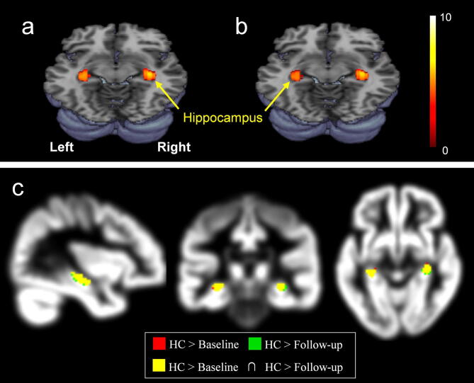
The efficacy of donepezil is well known for improving the cognitive performance in patients with mild cognitive impairment (MCI) and Alzheimer's disease (AD). Most of the recent neuroimaging studies focusing on the brain morphometry have dealt with the targeted brain structures, and thus it remains unknown how donepezil treatment influences the volume change over the whole brain areas including the cortical and subcortical regions and hippocampal subfields in particular. This study aimed to evaluate overall gray matter (GM) volume changes after donepezil treatment in MCI, which is a prodromal phase of AD, using voxel-based morphometry. Patients with MCI underwent the magnetic resonance imaging (MRI) before and after 6-month donepezil treatment. The cognitive function for MCI was evaluated using the questionnaires of the Korean version of the mini-mental state examination (K-MMSE) and Alzheimer's disease assessment scale-cognitive subscale (ADAS-Cog). Compared with healthy controls, patients with MCI showed significantly lower GM volumes in the hippocampus and its subfields, specifically in the right subiculum and left cornu ammonis (CA3). The average scores of K-MMSE in patients with MCI improved by 8% after donepezil treatment. Treated patients showed significantly higher GM volumes in the putamen, globus pailldus, and inferior frontal gyrus after donepezil treatment (p < 0.001). However, whole hippocampal volume in the patients decreased by 0.6% after 6-month treatment, and the rate of volume change in the left hippocampus was negatively correlated with the period of treatment. These findings will be useful for screening and tracking MCI, as well as understanding of the pathogenesis of MCI in connection with brain morphometric change.


Conflict of interest statement

The authors declare no competing interests.

Figures

Figure 1
Figure 1
3D atlas of the hippocampus and hippocampal subfield segmentation. CA, cornu ammonis. This figure was created using Freesurfer (version 6.0 https://surfer.nmr.mgh.harvard.edu) and Microsoft Powerpoint (version 16 https://www.microsoft.com).
Figure 2
Figure 2
Demonstration of the decreased gray matter volumes of the hippocampus: (a) untreated patients (baseline) relative to healthy control (HC), (b) treated patients (follow-up) relative to healthy controls (FWE corrected, p < 0.05), and (c) intersection set of {HC > baseline} and {HC > follow-up} which highlights the overlapping areas with different colors on the sagittal, coronal, and axial planes, i.e. green, HC > baseline; red, HC > follow-up; yellow, the intersection of the two sets, red and green. This figure was created using SPM (version 8 https://www.fil.ion.ucl.ac.uk/spm), MRIcron (version 6 https://www.nitrc.org/projects/mricron), and Microsoft Powerpoint (version 16 https://www.microsoft.com).
Figure 3
Figure 3
Brain regions with significantly increased GM volumes (a) and decreased GM volumes (b) in donepezil-treated patients compared with untreated patients [p < 0.001 (a) or p < 0.05 (b)]. IFG, inferior frontal gyrus. This figure was created using SPM (version 8 https://www.fil.ion.ucl.ac.uk/spm), MRIcron (version 6 https://www.nitrc.org/projects/mricron), and Microsoft Powerpoint (version 16 https://www.microsoft.com).
Figure 4
Figure 4
Comparison of the volumes of the hippocampal subfields in healthy controls, untreated patients (baseline), and treated patients (follow-up). Sub, subiculum; CA, cornu ammonis, PrS, presubiculum; ML, molecular layer of the hippocampus, GC-DG, granular cells layer of the dentate gyrus. This figure was created using SigmaPlot (version 13 https://systatsoftware.com/products/sigmaplot) and Microsoft Powerpoint (version 16 https://www.microsoft.com). *Significant difference (Bonferroni corrected, p < 0.05).
Figure 5
Figure 5
The rate of volume change in the left hippocampus is negatively correlated with the period of treatment in patients with MCI (Spearman’s rho = − 0.80, p = 0.005). Dotted lines show 95% CI. The rate of volume change between baseline and follow-up in MCI patients was calculated by the equation, (Volumefollow-up − Volumebaseline)/(Volumebaseline). This figure was created using SigmaPlot (version 13 https://systatsoftware.com/products/sigmaplot) and Microsoft Powerpoint (version 16 https://www.microsoft.com).

References

    1. Yuoksimaa E, et al. Modifying the minimum criteria for diagnosing amnestic MCI to improve prediction of brain atrophy and progression to Alzheimer's disease. Brain Imaging Behav. 2018 doi: 10.1007/s11682-018-0019-6. - DOI - PMC - PubMed
    1. Fumagalli F, Racagni G, Riva MA. The expanding role of BDNF: a therapeutic target for Alzheimer's disease? Pharmacogenomics. 2006;6:8–15. doi: 10.1038/sj.tpj.6500337. - DOI - PubMed
    1. Hashimoto M, et al. Does donepezil treatment slow the progression of hippocampal atrophy in patients with Alzheimer's disease? Am. J.. Psychiatry. 2005;162:676–682. doi: 10.1176/appi.ajp.162.4.676. - DOI - PubMed
    1. Stahl SM. The new cholinesterase inhibitors for Alzheimer's disease, Part 2: illustrating their mechanisms of action. J. Clin. Psychiatry. 2000;61:813–814. doi: 10.4088/JCP.v61n1101. - DOI - PubMed
    1. Modrego PJ, et al. Memantine versus donepezil in mild to moderate Alzheimer's disease: a randomized trial with magnetic resonance spectroscopy. Eur. J. Neurol. 2010;17:405–412. doi: 10.1111/j.1468-1331.2009.02816.x. - DOI - PubMed

Publication types

MeSH terms