

Breast Imaging Chameleon: Pseudoangiomatous Stromal Hyperplasia Presenting as Breast Malignancy
- PMID: 32617230
- PMCID: PMC7325414
- DOI: 10.7759/cureus.8359
Breast Imaging Chameleon: Pseudoangiomatous Stromal Hyperplasia Presenting as Breast Malignancy
Abstract
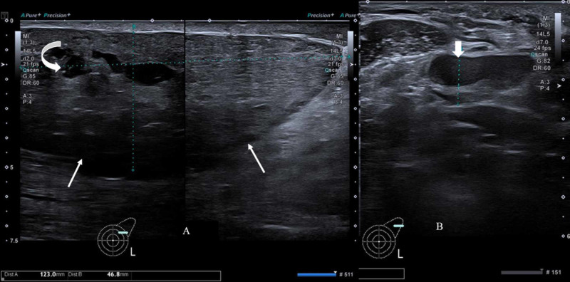
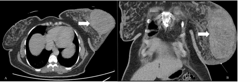
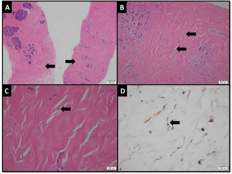
Pseudoangiomatous stromal hyperplasia (PASH) is a benign mesenchymal proliferative lesion of the breast, often an incidental finding on breast biopsy specimens and rarely presents as a palpable lump. The case being reported is interesting as a lactating female presented with gross left breast enlargement due to a huge firm mass with skin thickening and palpable left axillary lymph nodes. A provisional diagnosis of left breast malignancy was made and the patient extensively worked up with ultrasound, CT scan, bone scan and core biopsy. The histopathology, however, revealed PASH of the breast. There was no invasive or in situ malignancy. The patient was successfully managed conservatively.
Keywords: benign; breast; malignancy; palpable lump; pseudoangiomatous stromal hyperplasia; ultrasound.
Copyright © 2020, Raza et al.
Conflict of interest statement
The authors have declared that no competing interests exist.
Figures
References
-
- Pseudoangiomatous stromal hyperplasia: an overview. Virk RK, Khan A. https://pubmed.ncbi.nlm.nih.gov/20586640/ Arch Pathol Lab Med. 2010;134:1070–1074. - PubMed
-
- Pseudoangiomatous hyperplasia of mammary stroma. Vuitch MF, Rosen PP, Erlandson RA. Hum Pathol. 1986;17:185–191. - PubMed
-
- Pseudoangiomatous stromal hyperplasia (PASH) of the mammary gland: report of a case. Sasaki Y, Kamata S, Saito K, Nishikawa Y, Ogawa J. Surg Today. 2008;38:340–343. - PubMed
-
- Pseudoangiomatous stromal hyperplasia of the breast: multimodality review with pathologic correlation. Raj SD, Sahani VG, Adrada BE, et al. Curr Probl Diagno Radiol. 2017;46:130–135. - PubMed
-
- Pseudoangiomatous stromal hyperplasia: diagnosis, treatment and follow-up; description of a case-series. Donk WA, Oostenbroek RJ, Storm RK, Westenend PJ, Plaisier PW. https://benthamopen.com/contents/pdf/TOBCANJ/TOBCANJ-3-18.pdf Open Breast Cancer J. 2011;3:18–23.
Publication types
LinkOut - more resources
Full Text Sources