. 2020 Aug;51(4):421-435.
doi: 10.1007/s10735-020-09893-6. Epub 2020 Jul 2.

Advillin is a tuft cell marker in the mouse alimentary tract

Affiliations

Advillin is a tuft cell marker in the mouse alimentary tract

Anna-Lena Ruppert et al. J Mol Histol. 2020 Aug.

Abstract

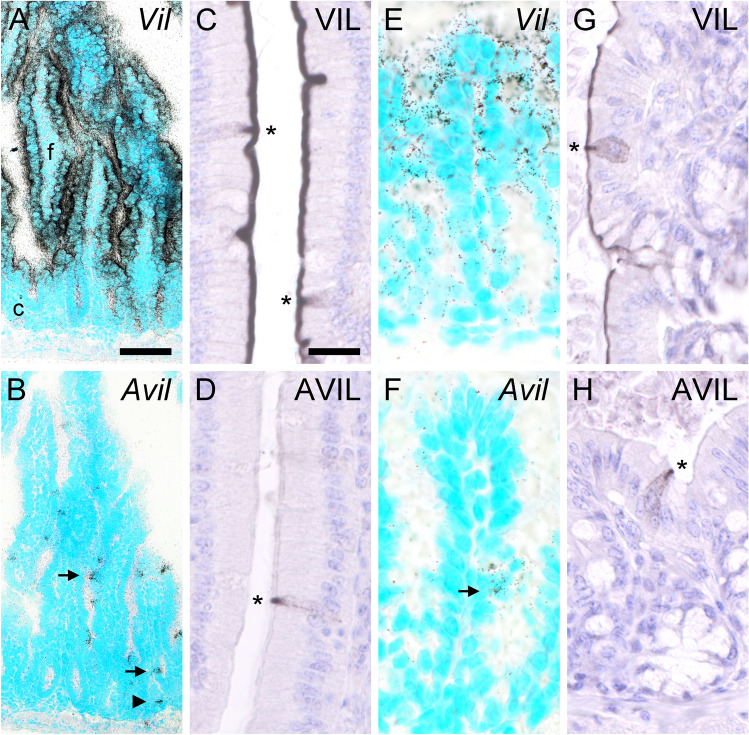
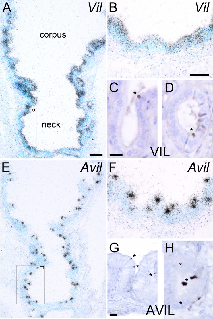
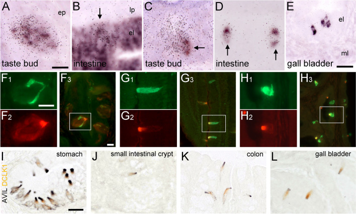
Tuft cells are a rare population of chemosensory cells at the mucosal surface epithelia of hollow organs. Their name-giving morphological feature is an apical tuft of stiff microvilli. Accordingly, the actin-binding protein, villin, was identified as one of the first tuft cell markers in immunohistochemical analysis. Unfortunately, villin expression is not restricted to tuft cells, but is also prominent e.g. in enterocytes, which limits the use of this gene as a marker and as an experimental tool to genetically target tuft cells. Here, we report that the villin-related protein, advillin, is a specific tuft cell marker in the gastro-intestinal and biliary tract epithelia. In situ hybridization and immunohistochemistry revealed that advillin expression, unlike villin, was restricted to solitary cholinergic tuft cells in the mucosal linings of the small and large intestine, and in the gall bladder. In the glandular stomach, villin and advillin mRNA were present in all epithelial cells, while detectable protein levels were confined to solitary tuft cells. Advillin expression was no longer detectable in the mucosa of the intestinal and biliary tract from Pou2f3 deficient mice that lack tuft cells. Finally, crossing Avil-Cre transgenic mice with a double-fluorescent reporter mouse line resulted in specific targeting of gastro-intestinal and biliary tuft cells. Our analysis introduces advillin as a selective marker and tool in histological and functional analysis of the alimentary tract tuft cell system.

Keywords: Biliary tract; Doublecortin-like kinase 1; Immunohistochemistry; In situ hybridization; Intestine; Villin.


Conflict of interest statement

The authors declare that they have no conflict of interest.

Figures

Fig. 1
Fig. 1
Villin and advillin expression patterns in mouse taste buds and in dorsal root ganglia. Vil mRNA (a), and Avil mRNA (b), where both detected with antisense (as) riboprobes in radioactive ISH in taste buds of the mouse vallate papilla. The use of sense (s) riboprobes for Vil (c) and Avil (d) did not result in specific staining. Villin-immunoreactivity outlined many, if not all taste cells, with the apical tip showing the most intense staining (e). Likewise, advillin-immunoreactive cells were present in taste buds, sometimes showing intense intra-cellular staining (f). Absence of villin-immunoreactivity (g), and presence of advillin-immunoreactivity (h) in dorsal root ganglion neurons. Sections from ISH were counter-stained with methyl green, sections from IHC with hemalaun. The bar in a represents 50 µm and applies to ad. The bar in e represents 20 µm and also applies to f. The bar in g represents 20 µm and also applies to h
Fig. 2
Fig. 2
Villin and advillin expression patterns in the mouse stomach. In the mouse stomach, the squamo-columnar junction (scj) represents the histological transition from the limiting ridge (lr) to the corpus (c). (a) Vil mRNA, detected by radioactive ISH, was present both in epithelial cells of the columnar aspect of the corpus, and in deeper glands. (b) Avil mRNA displayed a similar expression pattern, with no preference in staining intensity for individual cells. On the protein level, both villin- (c) and advillin- (d) immunoreactivities were confined to individual cells of the columnar epithelium (arrows) and in deeper glandular aspects (arrowheads). Especially the staining of the apical tips of the columnar and flask-shaped cells was most intense. Vil (e) and Avil (f) mRNA was present throughout the mucosal epithelium of the corpus, while immunoreactivity was confined to individual cells (arrows in g and h). Sections from ISH were counter-stained with methyl green, sections from IHC with hemalaun. The bar in a represents 20 µm and also applies to b. The bar in c represents 20 µm and also applies to d. The bar in e represents 50 µm and also applies to f. The bar in g represents 20 µm and also applies to h
Fig. 3
Fig. 3
Villin and advillin expression patterns in the mouse small and large intestine. (a) Vil mRNA, detected with antisense riboprobes in radioactive ISH, was present in the entire epithelial lining of the small intestine, shown here for duodenum. Note much stronger expression in folds (f) compared to crypts (c). (b) Avil mRNA was confined to individual epithelial cells, both in folds (arrow) and in crypts (arrowhead). (c) Villin-immunoreactivity was seen along the entire apical aspects of the duodenal mucosa, occasionally with additional cell body labeling (asterisks). (d) Advillin-immunoreactivity was confined to individual cells in the mucosal epithelium. Note prominent staining of the apical tip (asterisk). (e) In the large intestine, Vil mRNA was mainly present in the upper half of the crypts, shown here for the colon. (f) Again, presence of Avil mRNA was confined to individual cells. (g) Villin-immunoreactivity was seen along the entire apical aspects of the colonic mucosa, occasionally with additional cell body labeling (asterisk). (h) Advillin-immunoreactivity was confined to individual cells in the mucosal epithelium (asterisk). Sections from ISH were counter-stained with methyl green, sections from IHC with hemalaun. The bar in a represents 100 µm and also applies to b. The bar in c represents 20 µm and also applies to dh
Fig. 4
Fig. 4
Villin and advillin expression patterns in the mouse gall bladder. (a) Vil mRNA was present in the entire epithelial lining of the gall bladder, showing no regional accumulations e.g. in neck or corpus. (b) higher magnification view from the boxed area in a. (c) Villin-immunoreactivity was prominent in the apical tips of individual cells in the gall bladder main epithelium (asterisk), and (d) in associated glands (asterisk). (e) Avil mRNA was detected in individual cells in all parts of the gall bladder. (f) Higher magnification view from the boxed area in e. Advillin-immunoreactivity also was seen in individual cells along the inner lining of the gall bladder, both in the main epithelium (g) and in glands (h). Sections from ISH were counter-stained with methyl green, sections from IHC with hemalaun. The bar in a represents 100 µm and also applies to e. The bar in b represents 50 µm and also applies to f. The bar in c represents 10 µm and also applies to d and h. The bar in g represents 20 µm. (Color figure online)
Fig. 5
Fig. 5
Double-ISH and double-IHC experiments. Simultaneous detection of Vil (non-radioactive label) and Avil (radioactive label) mRNAs in a mouse taste bud (a), and in the duodenum (b). Note that Avil signals overlap with Vil (arrow in b). Simultaneous detection of Chat-Egfp (non-radioactive label) and Avil (radioactive label) mRNAs in a mouse taste bud (c), in the duodenum (d), and in the gall bladder (e). Note partial overlap in taste bud (arrow in c) and complete overlap of both labels in intestine (arrows) and in gall bladder. Double-IHC analysis of ChAT-EGFP (green label) with advillin (red label) in taste buds (f), in the duodenum (g), and in the gall bladder (h). The boxed area in the composite pictures f3, g3, and h3 is presented in magnification with the single label in f1, 2, g1, 2, and h1, 2. Double brightfield IHC analysis of advillin (black reaction product) with DCLK1 (brown reaction product) at the squamo-columnar junction of the stomach (i), in the small intestine (j), in the large intestine (k), and in the gall bladder (l). el epithelial layer; ep epidermis; lp lamina propria; ml muscle layer. The bar in a represents 20 µm and also applies to bd. The bar in e represents 25 µm. The bar in f1 represents 25 µm and also applies to f2, g1, 2, h1, 2. The bar in f3 represents 20 µm and also applies to g3 and h3. The bar in i represents 20 µm and also applies to jl. (Color figure online)
Fig. 6
Fig. 6
Advillin expression pattern in Pou2f3−/− mice. Avil mRNA, detected with antisense riboprobes in radioactive ISH, was present in vallate papilla taste buds (a), and in the epithelium of the glandular stomach (b) of Pou2f3−/− mice. Avil mRNA was absent from intestinal crypts and villi, but present in some neurons of the submucous (arrowhead) and myenteric (doubled arrowhead) plexus (c). Avil mRNA was absent from the gall bladder epithelium (d). Using IHC, advillin-immunoreactivity was detected faintly in taste buds (e, arrow), but was absent from the epithelium of stomach (f), duodenum (g), and gall bladder (h). Sections from ISH were counter-stained with methyl green, sections from IHC with hemalaun. c crypt; el epithelial layer; ep epidermis; lr limiting ridge; ml, muscle layer; v villus. The bars in a and b represent 100 µm. The bars in c and d represent 50 µm. The bar in e represents 20 µm and also applies to fh. (Color figure online)
Fig. 7
Fig. 7
Avil-Cre mice are a tool to genetically target tuft cells in the mouse alimentary tract. The mating of mice that harbor the double fluorescent reporter, mT/mG, with a mouse line that expressed Cre-recombinase under the control of the advillin promoter (Avil-Cre) resulted in cell-specific excision of mT due to flanking loxP (blue triangle) sites, and activation of Egfp expression (a). pA = polyadenylation site. Native EGFP fluorescence in fresh tissue representing Cre activity in vallate papilla (b), duodenal mucosa (c), and gall bladder (d). Enhancing EGFP detection with antibodies revealed presence of EGFP in vallate papilla taste buds, and in innervating presumably sensory nerve fibers (e). Similarly, EGFP was present in solitary cells at the limiting ridge (f1, marked by asterisk) and in the glandular corpus of the stomach (f2, marked by asterisk), and in nerve fibers (arrowhead in f2) traveling bottom-up in these glands. In the duodenum, solitary cells in the mucosal epithelium were labeled (asterisks) in addition to presumably sensory nerve fibers in the muscle layers (arrows in inset), while nerve cell bodies were not stained (arrowhead in inset) (g). EGFP-positive cells were present in the epithelium of the gall bladder (asterisks) (h). Double-immunofluorescence analysis of EGFP (i, green label) and advillin (k, red label) showed co-existence (k) in the duodenum. The bar in b represents 10 µm and also applies to d. The bar in c represents 20 µm. The bars in e, g (inset 10 µm), and h represent 50 µm. The bar in f represents 20 µm. The bar in i represents 20 µm and also applies to j, k. (Color figure online)

References

    1. Bezençon C, Fürholz A, Raymond F, Mansourian R, Métairon S, Le Coutre J, Damak S. Murine intestinal cells expressing Trpm5 are mostly brush cells and express markers of neuronal and inflammatory cells. J Comp Neurol. 2008;509:514–525. doi: 10.1002/cne.21768. - DOI - PubMed
    1. Bezençon C, le Coutre J, Damak S. Taste-signaling proteins are coexpressed in solitary intestinal epithelial cells. Chem Senses. 2007;32:41–49. doi: 10.1093/chemse/bjl034. - DOI - PubMed
    1. Bjerknes M, et al. Origin of the brush cell lineage in the mouse intestinal epithelium. Dev Biol. 2012;362:194–218. doi: 10.1016/j.ydbio.2011.12.009. - DOI - PMC - PubMed
    1. Chuang YC, Lee CH, Sun WH, Chen CC. Involvement of advillin in somatosensory neuron subtype-specific axon regeneration and neuropathic pain. Proc Natl Acad Sci USA. 2018;115:E8557–E8566. doi: 10.1073/pnas.1716470115. - DOI - PMC - PubMed
    1. Deckmann K, et al. Bitter triggers acetylcholine release from polymodal urethral chemosensory cells and bladder reflexes. Proc Natl Acad Sci USA. 2014;111:8287–8292. doi: 10.1073/pnas.1402436111. - DOI - PMC - PubMed

LinkOut - more resources