

The Leaf Extracts of Toona sinensis and Fermented Culture Broths of Antrodia camphorata Synergistically Cause Apoptotic Cell Death in Promyelocytic Leukemia Cells
- PMID: 32618215
- PMCID: PMC7336824
- DOI: 10.1177/1534735420923734
The Leaf Extracts of Toona sinensis and Fermented Culture Broths of Antrodia camphorata Synergistically Cause Apoptotic Cell Death in Promyelocytic Leukemia Cells
Abstract
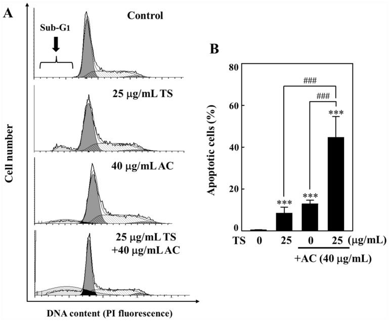
Toona sinensis is a common edible vegetable that is used in certain Chinese dishes and has importance in folk medicine. The leaf extracts of T sinensis possess and exhibit anticancer efficacy against various cancer cell types. In Taiwanese folklore, Antrodia camphorata, also known as "Niu-Cheng-Zi," is used in traditional medicine to treat various illnesses. Its fruit and mycelium possess various potent antiproliferative properties. Two studies from our group have reported that T sinensis or A camphorata has the ability to cause apoptosis in various cancer cells. Conversely, underlying molecular mechanisms and any beneficial effects remain unknown. This study shows anticancer efficacy for both T sinensis and A camphorata co-treatments that target HL-60 cells. The combination index values indicate that 40 µg/mL of T sinensis and 25 µg/mL of A camphorata as a combined treatment shows a synergetic effect, which reduces HL-60 cell proliferation. Alternately, this treatment exhibited no cytotoxic effects for human umbilical vein endothelial cells. Western blot data showed that T sinensis and A camphorata as a combined treatment result in augmented expression of apoptosis, cytochrome c release, Bcl-2 inhibition, expression of Bax, Fas, and FasL, as well as the cleavage of Bid in HL-60 cells. Moreover, this combined treatment overshadowed monotherapy in its ability to inhibit uPAR, MMP-9, MMP-2, COX-2 expression, and PGE2 secretions. Our study strongly implies that this combined treatment offers more beneficial effects to suppress and treat leukemia due to apoptosis-mediated cell inhibition. Further in vivo studies related to the combined treatment could establish its future potential.
Keywords: Antrodia camphorate; COX-2; HL-60 cells; MMP; Toona sinensis; apoptosis; leukemia.
Conflict of interest statement
Figures


Similar articles
-
Induction of apoptosis by Antrodia camphorata in human premyelocytic leukemia HL-60 cells.Nutr Cancer. 2004;48(2):189-97. doi: 10.1207/s15327914nc4802_9. Nutr Cancer. 2004. PMID: 15231454
-
Toona sinensis extracts induces apoptosis via reactive oxygen species in human premyelocytic leukemia cells.Food Chem Toxicol. 2006 Dec;44(12):1978-88. doi: 10.1016/j.fct.2006.06.027. Epub 2006 Jul 18. Food Chem Toxicol. 2006. PMID: 16945458
-
Growth inhibition and induction of apoptosis in MCF-7 breast cancer cells by Antrodia camphorata.Cancer Lett. 2006 Jan 18;231(2):215-27. doi: 10.1016/j.canlet.2005.02.004. Cancer Lett. 2006. PMID: 16399223
-
Review of biological and pharmacological activities of the endemic Taiwanese bitter medicinal mushroom, Antrodia camphorata (M. Zang et C. H. Su) Sh. H. Wu et al. (higher Basidiomycetes).Int J Med Mushrooms. 2012;14(3):241-56. doi: 10.1615/intjmedmushr.v14.i3.20. Int J Med Mushrooms. 2012. PMID: 22577975 Review.
-
Niuchangchih (Antrodia camphorata) and its potential in treating liver diseases.J Ethnopharmacol. 2009 Jan 21;121(2):194-212. doi: 10.1016/j.jep.2008.10.039. Epub 2008 Nov 17. J Ethnopharmacol. 2009. PMID: 19061947 Review.
Cited by
-
Anti-Aging Potential of Plants of the Anak Dalam Tribe, Jambi, Indonesia.Pharmaceuticals (Basel). 2023 Sep 14;16(9):1300. doi: 10.3390/ph16091300. Pharmaceuticals (Basel). 2023. PMID: 37765107 Free PMC article. Review.
-
Mitochondrial dysfunction and cell death induced by Toona sinensis leaf extracts through MEK/ERK signaling in glioblastoma cells.PLoS One. 2025 May 9;20(5):e0320849. doi: 10.1371/journal.pone.0320849. eCollection 2025. PLoS One. 2025. PMID: 40343958 Free PMC article.
References
-
- Robak T, Wierzbowska A. Current and emerging therapies for acute myeloid leukemia. Clin Ther. 2009;31(pt 2):2349-2370. - PubMed
-
- Bold RJ, Termuhlen PM, McConkey DJ. Apoptosis, cancer and cancer therapy. Surg Oncol. 1997;6:133-142. - PubMed
-
- Kamesaki H. Mechanisms involved in chemotherapy-induced apoptosis and their implications in cancer chemotherapy. Int J Hematol. 1998;68:29-43. - PubMed
Publication types
MeSH terms
Substances
Supplementary concepts
LinkOut - more resources
Full Text Sources
Medical
Research Materials
Miscellaneous
