

An Atypical Case of Autochthonous Cutaneous Leishmaniasis Associated with Naturally Infected Phlebotomine Sand Flies in Texas, United States
- PMID: 32618254
- PMCID: PMC7543804
- DOI: 10.4269/ajtmh.20-0107
An Atypical Case of Autochthonous Cutaneous Leishmaniasis Associated with Naturally Infected Phlebotomine Sand Flies in Texas, United States
Abstract
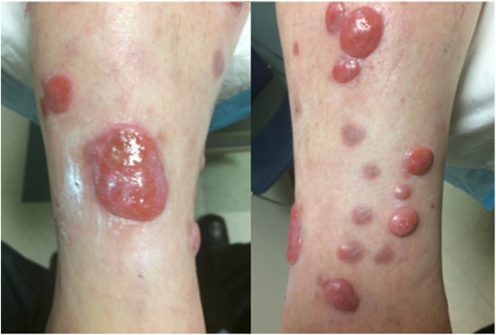
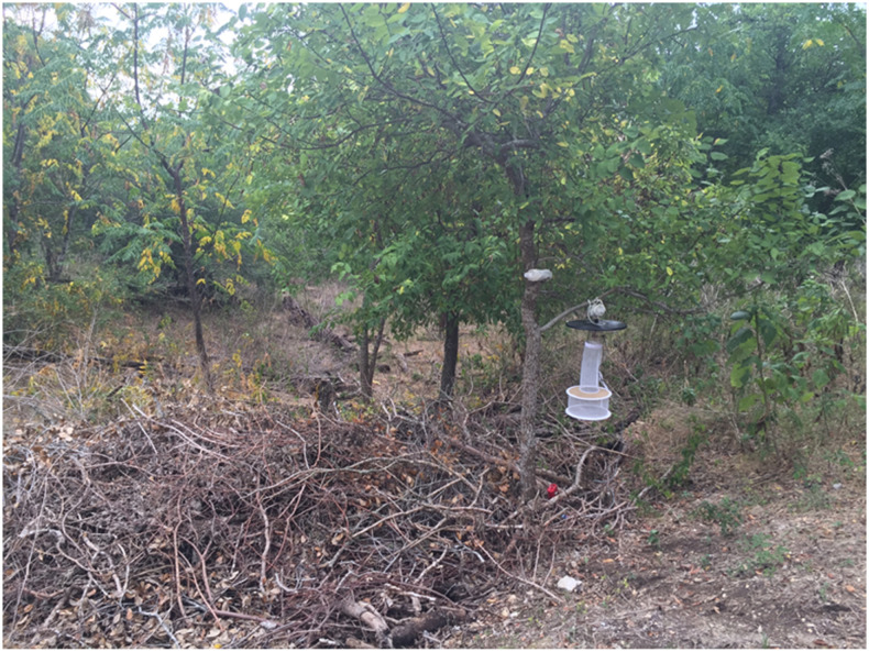
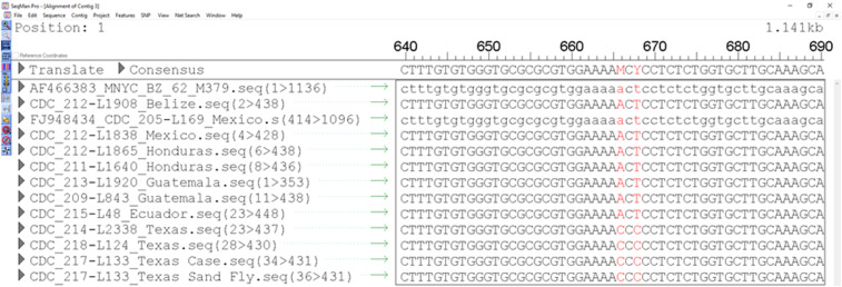
In the United States, phlebotomine sand flies carrying Leishmania (Leishmania) mexicana are endemic along the southern border. However, relatively little is known about the enzootic and zoonotic transmission of L. (L.) mexicana within the United States, and autochthonous cases of the consequent disease are rarely reported. We investigated an atypical case of cutaneous leishmaniasis (CL) caused by L. (L.) mexicana in a patient from central Texas which did not respond to a typical antileishmanial chemotherapy. We also investigated sand fly vectors around the patient's residence. PCR followed by DNA sequencing was used for determination of Leishmania spp., sand fly species, and host blood meal source. The L. (L.) mexicana genotype from the patient was identical to one found in a positive sand fly. Moreover, this genotype presented the same single-nucleotide polymorphisms as other historical CL cases acquired in Texas over the last 10 years, but distinct from those originating in Mexico and Central America. Three sand fly species were identified among the samples analyzed (n = 194), the majority of which were Lutzomyia (Dampfomyia) anthophora (n = 190), of which four specimens tested positive for Leishmania and two blood-fed specimens showed the presence of a human blood meal. This study highlights the complexity of clinical management of CL in a setting where the disease is infrequently encountered. The detection of human blood in Lu. (D.) anthophora is the first documentation of anthropophagy in this species. This is the first report of wild-caught, naturally infected sand flies found in association with an autochthonous case of human leishmaniasis and the specific strain of Leishmania (Leishmania) mexicana in the United States.
Conflict of interest statement
Disclaimer: The findings and conclusions in this article are those of the authors and do not necessarily represent the official position of the CDC.
Figures

References
-
- Herwaldt BL, 1999. Leishmaniasis. Lancet 354: 1191–1199. - PubMed
-
- Desjeux P, 2004. Leishmaniasis: current situation and new perspectives. Comp Immunol Microbiol Infect Dis 27: 305–318. - PubMed
-
- Reithinger R, Dujardin J-C, Louzir H, Pirmez C, Alexander B, Brooker S, 2007. Cutaneous leishmaniasis. Lancet Infect Dis 7: 581–596. - PubMed
-
- Bailey MS, Lockwood DNJ, 2007. Cutaneous leishmaniasis. Clin Dermatol 25: 203–211. - PubMed
Publication types
MeSH terms
LinkOut - more resources
Full Text Sources
Miscellaneous
