. 2020 Jul;10(3):e124.
doi: 10.1002/ctm2.124. Epub 2020 Jul 3.

Osteocrin attenuates inflammation, oxidative stress, apoptosis, and cardiac dysfunction in doxorubicin-induced cardiotoxicity

Affiliations

Osteocrin attenuates inflammation, oxidative stress, apoptosis, and cardiac dysfunction in doxorubicin-induced cardiotoxicity

Can Hu et al. Clin Transl Med. 2020 Jul.

Abstract

Background: Inflammation, oxidative stress, and apoptosis contribute to the evolution of doxorubicin (DOX)-induced cardiotoxicity. Osteocrin (OSTN) is a novel secretory peptide mainly derived from the bone and skeletal muscle, and plays critical roles in regulating bone growth and physical endurance. Inspiringly, OSTN was also reported to be abundant in the myocardium that functioned as a therapeutic agent against cardiac rupture and congestive heart failure in mice after myocardial infarction. Herein, we investigated the role and potential mechanism of OSTN in DOX-induced cardiotoxicity.

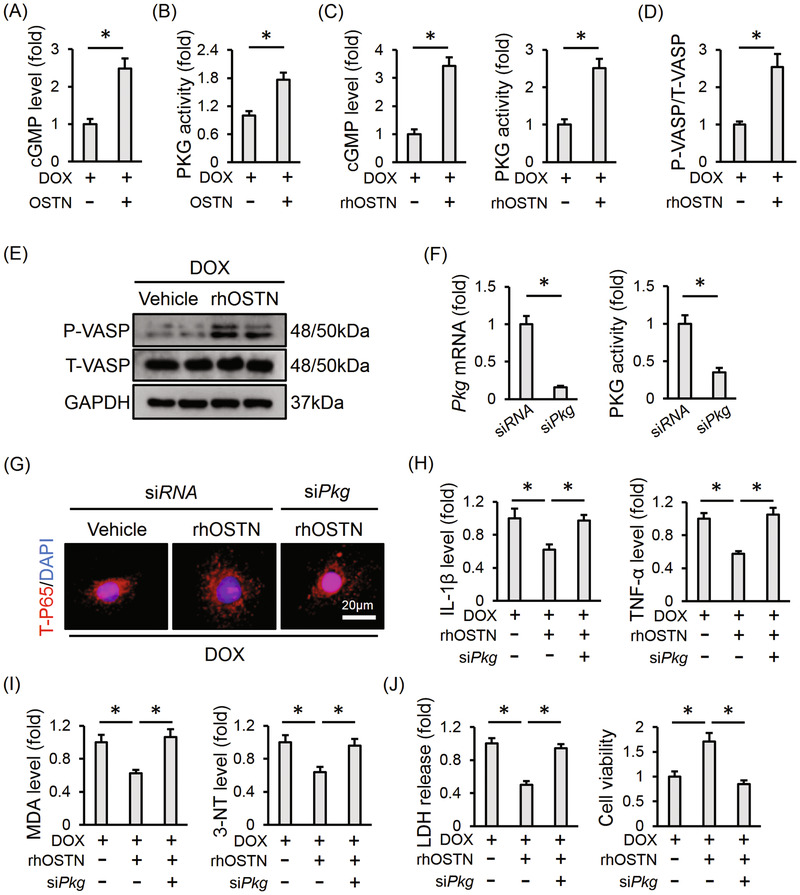
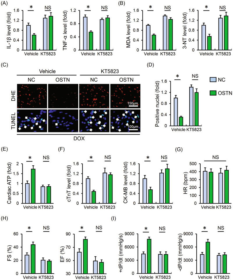
Methods: Cardiac-restrict OSTN overexpression was performed by the intravenous injection of a cardiotropic AAV9 vector, and subsequently the mice received 15 mg/kg DOX injection (i.p., once) to induce acute cardiac injury. Besides, H9C2 cell lines were used to assess the possible role of OSTN in vitro by incubating with recombinant human OSTN or small interfering RNA against Ostn (siOstn). To clarify the involvement of protein kinase G (PKG), KT5823 and siPkg were used in vivo and in vitro. Mice were also administrated intraperitoneally with 5 mg/kg DOX weekly for consecutive 3 weeks at a cumulative dose of 15 mg/kg to mimic the cardiotoxic effects upon chronic DOX exposure.

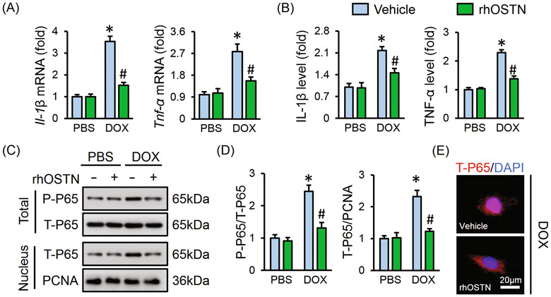
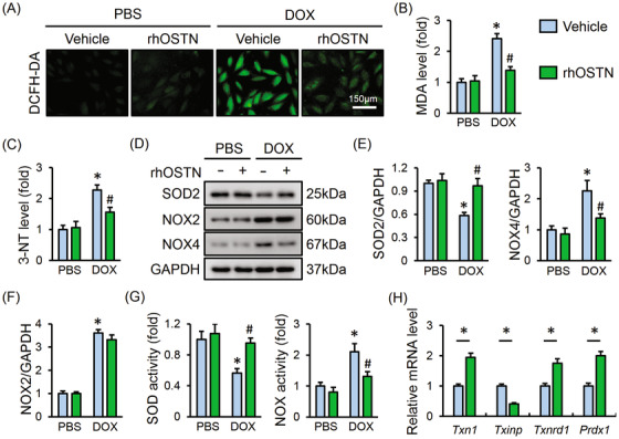
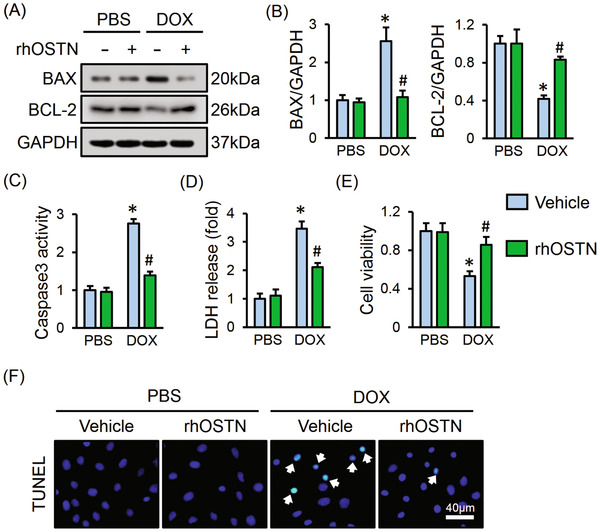
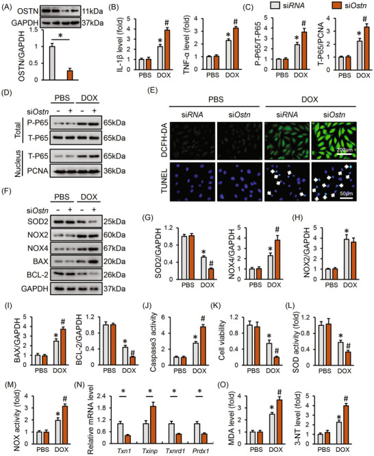
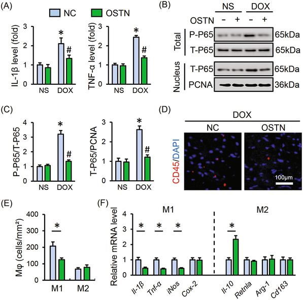
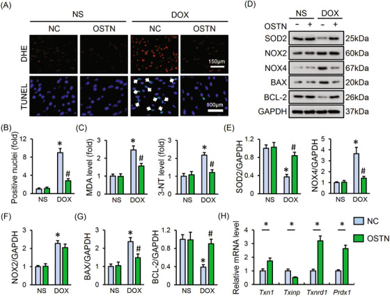
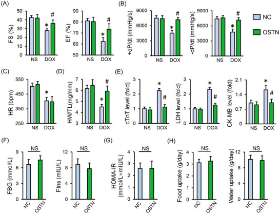
Results: OSTN treatment notably attenuated, whereas OSTN silence exacerbated inflammation, oxidative stress, and cardiomyocyte apoptosis in DOX-treated H9C2 cells. Besides, cardiac-restrict OSTN-overexpressed mice showed an alleviated cardiac injury and malfunction upon DOX injection. Mechanistically, we found that OSTN activated PKG, while PKG inhibition abrogated the beneficial effect of OSTN in vivo and in vitro. As expected, OSTN overexpression also improved cardiac function and survival rate in mice after chronic DOX treatment.

Conclusions: OSTN protects against DOX-elicited inflammation, oxidative stress, apoptosis, and cardiac dysfunction via activating PKG, and cardiac gene therapy with OSTN provides a novel therapeutic strategy against DOX-induced cardiotoxicity.

Keywords: apoptosis; doxorubicin; inflammation; osteocrin; oxidative stress.


Conflict of interest statement

The authors declare no conflict of interest.

Figures

FIGURE 1
FIGURE 1
OSTN alleviates DOX‐induced inflammation in vitro. H9C2 cells were treated with rhOSTN (5 µg/mL) or an equal volume of vehicle as the control in the presence or absence of DOX (1 µmol/L) for 24 h. A, The mRNA levels of inflammatory markers, Il‐1β and Tnf‐α (n = 6). B, The releases of IL‐1β, TNF‐α from H9C2 cells to the medium (n = 6). C and D, Representative western blot images and the quantitative results (n = 6). E, Immunofluorescence staining of T‐P65 in H9C2 cells (n = 6). Values represent the mean ± SD. *< .05 versus PBS + Vehicle; # < .05 versus DOX + Vehicle
FIGURE 2
FIGURE 2
OSTN attenuates DOX‐induced oxidative stress in vitro. A, Representative images of DCFH‐DA staining in H9C2 cells treated with rhOSTN in the presence or absence of DOX (n = 6). B and C, The level of MDA and 3‐NT in H9C2 cells (n = 8). D‐F, Representative western blot images and the corresponding statistical results (n = 6). G, Total SOD activity and NOX activity in H9C2 cells (n = 8). H, The mRNA level of Txn1, Txinp, Txnrd1, and Prdx1 in DOX‐treated H9C2 cells with or without rhOSTN protection (n = 6). Values represent the mean ± SD. * < .05 versus PBS + Vehicle; # < .05 versus DOX + Vehicle. In Figure 2H, * < .05 versus the matched group
FIGURE 3
FIGURE 3
OSTN protects against DOX‐induced cardiomyocyte apoptosis in vitro. A and B, Representative western blot images and the corresponding statistical results (n = 6). C, Caspase3 activity in H9C2 cells (n = 6). D, LDH release from H9C2 cells (n = 6). E, Cell viability assessed by CCK‐8 kits (n = 8). F, Representative images of TUNEL staining in H9C2 cells (n = 6). Values represent the mean ± SD. * < .05 versus PBS + Vehicle; # < .05 versus DOX + Vehicle
FIGURE 4
FIGURE 4
Ostn deficiency exacerbates DOX‐induced inflammation, oxidative stress and apoptosis in vitro. H9C2 cells were pre‐infected with siOstn (50 nmol/L) or siRNA (50 nmol/L) for 4 h and then maintained in normal medium for 24 h before DOX (1 µmol/L) incubation for additional 24 h. A, The efficiency of siOstn in H9C2 cells (n = 6). B, The IL‐1β and TNF‐α levels in the medium released from H9C2 cells (n = 6). C and D, Representative western blot images and the corresponding statistical results (n = 6). E, Representative images of DCFH‐DA and TUNEL stating (n = 6). F‐I, Representative western blot images and the corresponding statistical results (n = 6). J, Caspase3 activity in H9C2 cells (n = 8). K, Cell viability assessed by CCK‐8 kits (n = 8). L and M, SOD and NOX activity in H9C2 cells (n = 8). N, The mRNA level of Txn1, Txinp, Txnrd1, and Prdx1 in H9C2 cells (n = 6). O, The level of MDA, 3‐NT in H9C2 cells (n = 8). Values represent the mean ± SD. * < .05 versus PBS + siRNA; # < .05 versus DOX + siRNA. In Figure 4A,N, * < .05 versus the matched group
FIGURE 5
FIGURE 5
OSTN suppresses DOX‐induced inflammatory response in vivo. Mice were exposed to a single intravenous injection of AAV9‐OSTN (OSTN) or AAV9‐NC (NC) at a dosage of 1 × 10¹¹ viral genome per mouse and then maintained for 4 weeks, followed by a single intraperitoneal injection of DOX (15 mg/kg) for 8 days to generate DOX‐induced acute cardiotoxicity in mice. A, The IL‐1β and TNF‐α levels in myocardial tissues (n = 6). B and C, Representative western blot images and the corresponding statistical results (n = 6). (D) Representative images of CD45 staining in heart tissues (n = 6). E, Statistical results of M1‐type and M2‐type macrophages infiltration to the myocardium (n = 6). F, The mRNA level of M1‐type macrophage markers (Il‐1β, Tnf‐α, iNos, and Cox‐2), M2‐type macrophage markers (Il‐10, Retnla, Arg‐1, and Cd163) in murine hearts (n = 6). Values represent the mean ± SD. * < .05 versus NS + NC, # < .05 versus DOX+NC. In Figure 4E,F, * < .05 versus the matched group
FIGURE 6
FIGURE 6
OSTN blocks DOX‐induced oxidative stress and cardiomyocyte apoptosis in vivo. A, Representative images of DHE and TUNEL staining in myocardium (n = 8). B, Quantitative results of TUNEL‐positive nuclei in heart tissues (n = 8). C, The level of MDA, 3‐NT in heart tissues (n = 6). D‐G, Representative western blot images and the corresponding statistical results (n = 6). H, The mRNA level of Txn1, Txinp, Txnrd1, and Prdx1 in heart tissues (n = 6). Values represent the mean ± SD. * < .05 versus NS + NC; # < 0.05 versus DOX + NC. In Figure 6H, * < .05 versus the matched group
FIGURE 7
FIGURE 7
OSTN improves DOX‐induced acute cardiotoxicity in mice. A and B, Cardiac functional parameters of fractional shortening (FS), ejection fraction (EF), and the peak rates of isovolumic pressure development and pressure decay (±dP/dt) in left ventricles (n = 8). C, Heart rate (HR) (n = 8). D, Heart weight to tibial length ratio (HW/TL) (n = 8). E, Serum levels of cTnT, LDH, and CK‐MB in mice with or without OSTN overexpression after DOX injection (n = 8). F, Levels of fasting blood glucose (FBG) and fasting serum insulin (FIns) in control mice with or without OSTN overexpression (n = 8). G, The homeostasis model of assessment for insulin resistance (HOMA‐IR) index (n = 8). H, Food uptake and water uptake in control mice with or without OSTN overexpression (n = 10). Values represent the mean ± SD. * < .05 versus NS+NC; # < .05 versus DOX + NC; NS, no significance
FIGURE 8
FIGURE 8
OSTN prevents DOX‐induced inflammation, oxidative stress, and apoptosis via activating PKG. A, Myocardial cGMP level with or without OSTN overexpression after DOX injection (n = 6). B, Relative PKG activity in the myocardium (n = 6). C, Relative cGMP level and PKG activity in DOX‐treated H9C2 cells with or without rhOSTN incubation for 24 h (n = 6). D and E, Representative western blot images and the corresponding statistical results in H9C2 cells with or without rhOSTN incubation (n = 6). F, H9C2 cells were incubated with siPkg (50 nmol/L) or siRNA (50 nmol/L) for 4 h and then maintained in normal medium for additional 24 h. The efficiency of siPkg in H9C2 cells was detected by western blot (n = 6). G, H9C2 cells were pre‐infected with siPkg or siRNA, and then received DOX insult (1µmol/L) with or without rhOSTN protection (5 µg/mL). Immunofluorescence staining of T‐P65 in H9C2 cells (n = 6). H, The releases of IL‐1β and TNF‐α from H9C2 cells to the medium (n = 6). I, The levels of MDA and 3‐NT in H9C2 cells (n = 6). J, LDH release from H9C2 cells and the quantitative data of cell viability (n = 6). Values represent the mean ± SD. * < .05 versus the matched group
FIGURE 9
FIGURE 9
PKG inhibition abolishes the beneficial effect of OSTN in vivo. After AAV9 treatment, mice were intraperitoneally injected with KT5823 (1 mg/kg/day) or an equal volume of vehicle for consecutive 3 days prior to DOX treatment. A, The IL‐1β and TNF‐α levels in myocardial tissues (n = 6). B, The level of MDA and 3‐NT in heart samples (n = 6). C, Representative images of DHE and TUNEL staining in the myocardium (n = 8). D, Quantitative results of TUNEL‐positive nuclei in heart tissues (n = 8). E, Cardiac adenosine triphosphate (ATP) level in mice (n = 6). F, Serum levels of cTnT and CK‐MB in mice (n = 6). G, HR in mice among groups (n = 8). H and I, The statistical data of FS, EF, and ±dP/dt (n = 8). Values represent the mean ± SD. * < .05 versus the matched group; NS, no significance

References

    1. Silber JH, Barber G. Doxorubicin‐induced cardiotoxicity. N Engl J Med. 1995;333:1359‐1360. - PubMed
    1. Hutchinson L. Targeted therapies: doxorubicin and sorafenib improves survival in patients with advanced hepatocellular carcinoma. Nat Rev Clin Oncol. 2011;8:61. - PubMed
    1. Lu J, Li J, Hu Y, et al. Chrysophanol protects against doxorubicin‐induced cardiotoxicity by suppressing cellular PARylation. Acta Pharm Sin B. 2019;9:782‐793. - PMC - PubMed
    1. Tebbi CK, London WB, Friedman D, et al. Dexrazoxane‐associated risk for acute myeloid leukemia/myelodysplastic syndrome and other secondary malignancies in pediatric Hodgkin's disease. J Clin Oncol. 2007;25:493‐500. - PubMed
    1. Zhang X, Hu C, Kong CY, et al. FNDC5 alleviates oxidative stress and cardiomyocyte apoptosis in doxorubicin‐induced cardiotoxicity via activating AKT. Cell Death Differ. 2020;27:540‐555. - PMC - PubMed

LinkOut - more resources