. 2021 Feb 16;27(3):388-406.
doi: 10.1093/ibd/izaa161.

Inhibition of APE1/Ref-1 Redox Signaling Alleviates Intestinal Dysfunction and Damage to Myenteric Neurons in a Mouse Model of Spontaneous Chronic Colitis

Affiliations

Inhibition of APE1/Ref-1 Redox Signaling Alleviates Intestinal Dysfunction and Damage to Myenteric Neurons in a Mouse Model of Spontaneous Chronic Colitis

Lauren Sahakian et al. Inflamm Bowel Dis. .

Abstract

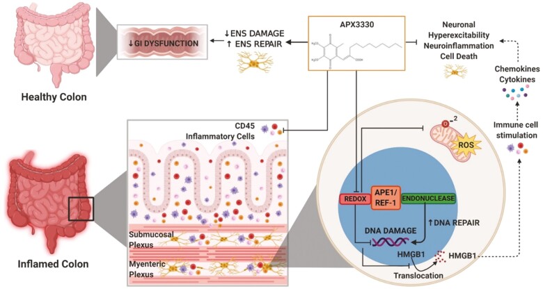
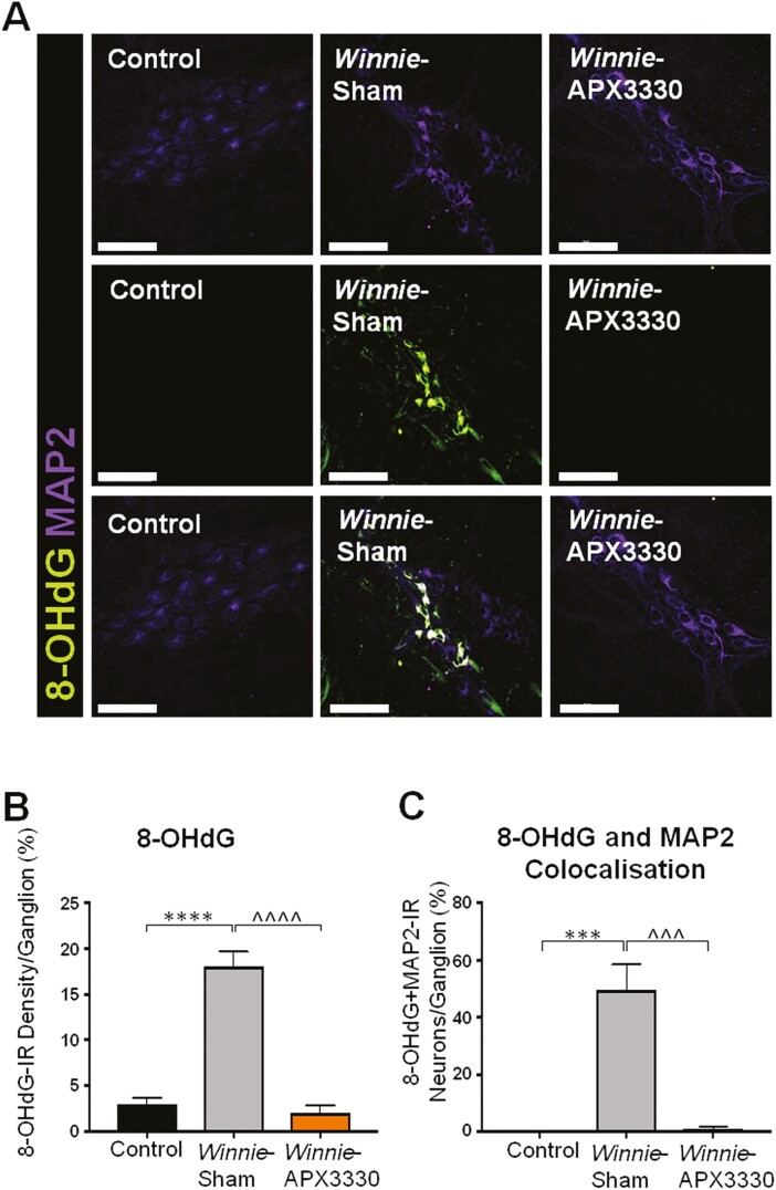
Background: Inflammatory bowel disease (IBD) associates with damage to the enteric nervous system (ENS), leading to gastrointestinal (GI) dysfunction. Oxidative stress is important for the pathophysiology of inflammation-induced enteric neuropathy and GI dysfunction. Apurinic/apyrimidinic endonuclease 1/redox factor-1 (APE1/Ref-1) is a dual functioning protein that is an essential regulator of the cellular response to oxidative stress. In this study, we aimed to determine whether an APE1/Ref-1 redox domain inhibitor, APX3330, alleviates inflammation-induced oxidative stress that leads to enteric neuropathy in the Winnie murine model of spontaneous chronic colitis.

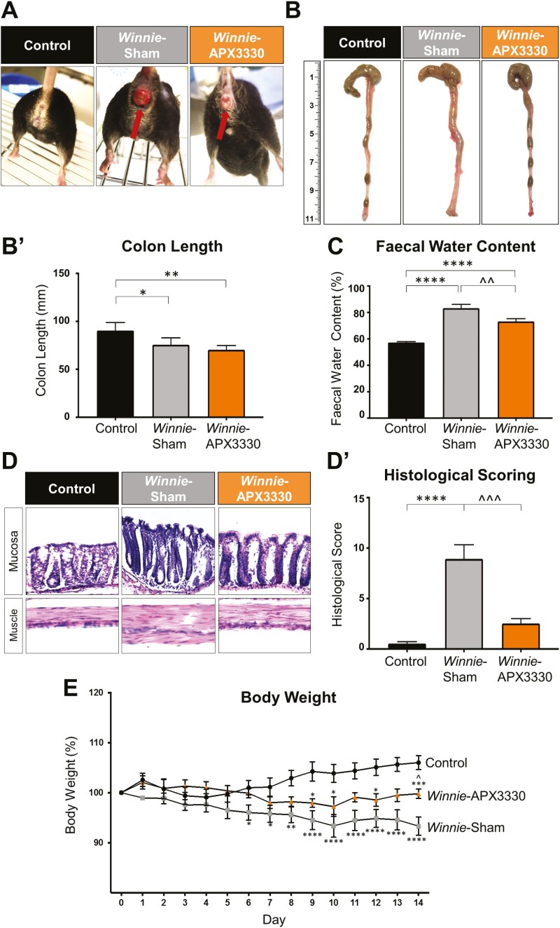
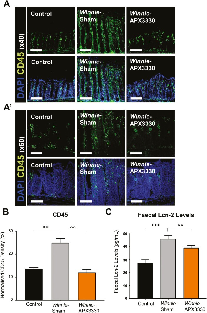
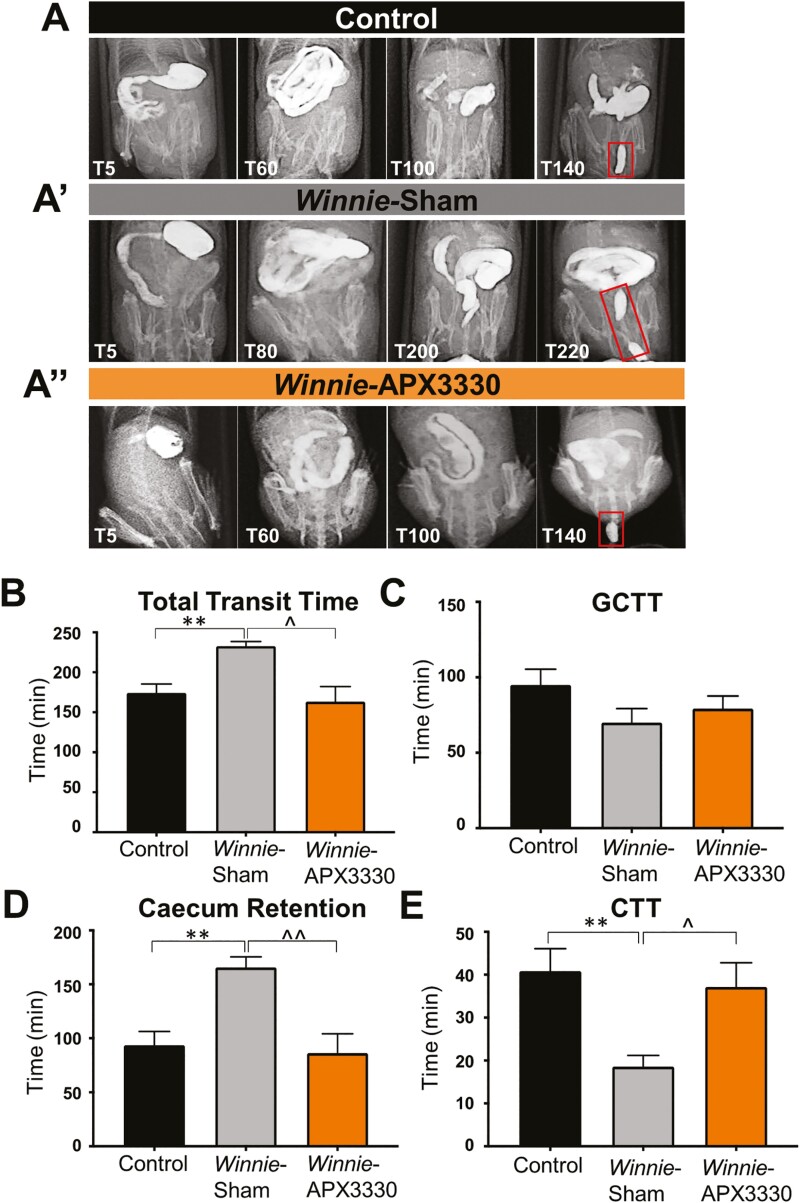
Methods: Winnie mice received APX3330 or vehicle via intraperitoneal injections over 2 weeks and were compared with C57BL/6 controls. In vivo disease activity and GI transit were evaluated. Ex vivo experiments were performed to assess functional parameters of colonic motility, immune cell infiltration, and changes to the ENS.

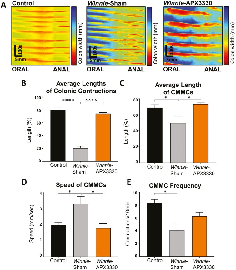
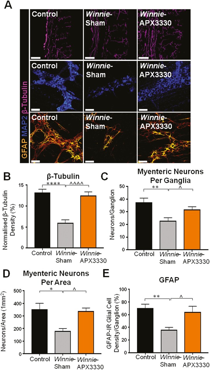
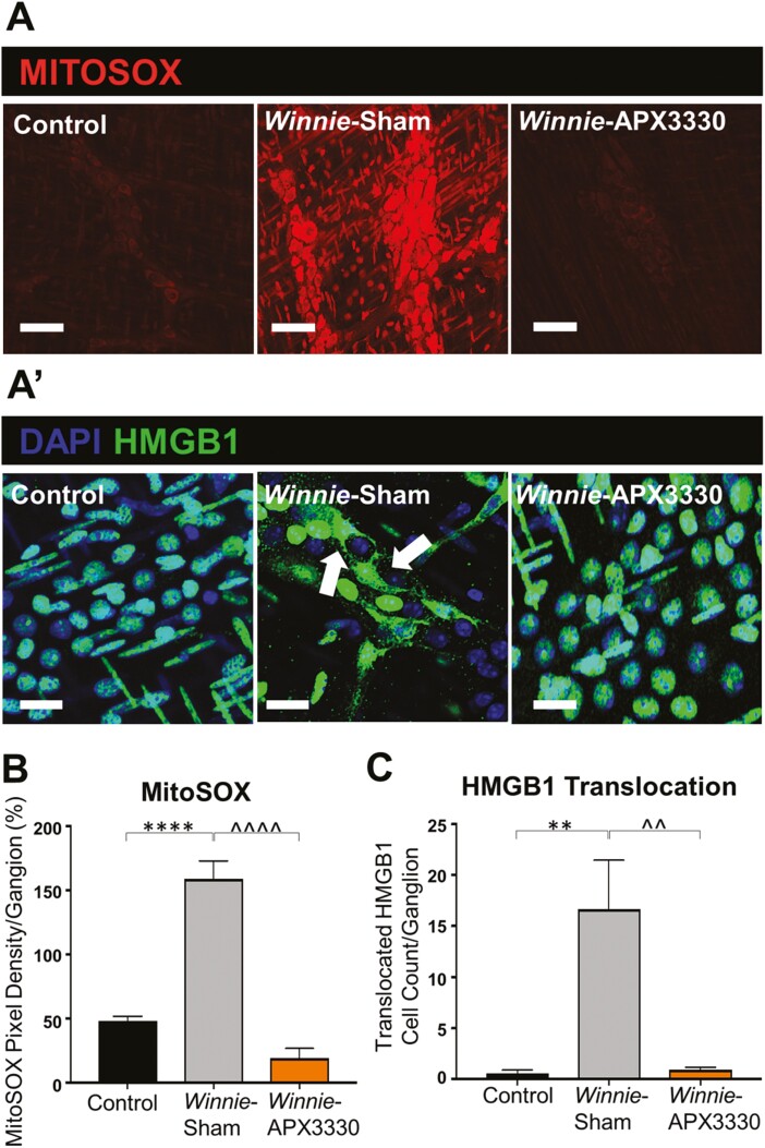
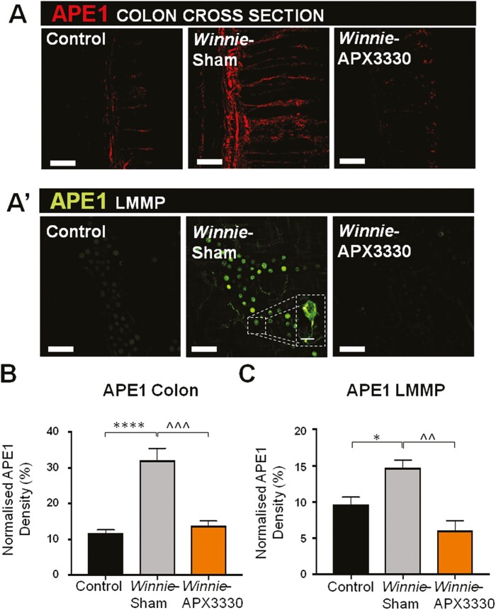
Results: Targeting APE1/Ref-1 redox activity with APX3330 improved disease severity, reduced immune cell infiltration, restored GI function ,and provided neuroprotective effects to the enteric nervous system. Inhibition of APE1/Ref-1 redox signaling leading to reduced mitochondrial superoxide production, oxidative DNA damage, and translocation of high mobility group box 1 protein (HMGB1) was involved in neuroprotective effects of APX3330 in enteric neurons.

Conclusions: This study is the first to investigate inhibition of APE1/Ref-1's redox activity via APX3330 in an animal model of chronic intestinal inflammation. Inhibition of the redox function of APE1/Ref-1 is a novel strategy that might lead to a possible application of APX3330 for the treatment of IBD.

Keywords: APE1/Ref-1; APX3330; DNA damage; IBD; chronic intestinal inflammation; enteric nervous system; oxidative stress.


Figures

Graphical Abstract
Graphical Abstract
APX3330 treatment in the animal model of chronic spontaneous colitis inhibits APE1/Ref-1 redox signalling, mitochondrial superoxide production, oxidative DNA damage and translocation of HMGB1 from the nucleus into cytoplasm and extracellularly. Extracellular HMGB1 stimulates immune cells to release cytokines and chemokines. APX3330 treatment alleviates the immune response. Suppressing APE1/Ref-1 redox activity enhances its endonuclease repair activity. These mechanisms alleviate damage to the enteric nervous system and GI dysfunction leading to improved clinical signs associated with chronic colitis.
FIGURE 1.
FIGURE 1.
The effects of APX3330 treatment on clinical symptoms in Winnie mice. A, Images of a C57BL/6 control mouse and a Winnie sham-treated mouse with severe intestinal inflammation and rectal prolapse with blood vessel proliferation and edema before and after 14 days of APX3330 treatment. B, B’. Colons from C57BL/6 control, Winnie sham-treated and Winnie APX3330-treated mice were excised and their length (mm) measured at day 15 (n = 5/group). C, Fecal water content (the difference between the wet and dry weight) was calculated as a percent of the wet weight of fresh fecal pellets measured at day 14 of treatment (n = 7/group). D, D’. Gross morphology of the distal colon was assessed by H&E staining and histological scoring was quantified in distal colon cross-sections from C57BL/6 control, Winnie sham-treated and Winnie APX3330-treated mice (n = 5/group). E, Body weight loss or gain in C57BL/6 control (n = 10), Winnie sham-treated (n = 10) and Winnie APX3330-treated (n = 8) mice over the 14-day period. Data expressed as mean ± SEM, *P < 0.05, **P < 0.01, ***P < 0.001, ****P < 0.0001 compared with C57BL/6 control mice; ^P < 0.05, ^^P < 0.01 compared with Winnie sham-treated mice.
FIGURE 2.
FIGURE 2.
The anti-inflammatory effects of APX3330 treatment in the colon of Winnie mice. A, and A’) CD45+ leukocytes were labeled using a leukocyte marker anti-CD45+ (green) antibody in the colon cross-sections. Mucosal epithelial cells are labeled with nuclei marker DAPI (blue; A: scale bar = 50 µm, 40x magnification; A’: scale bar = 30 µm, 60x magnification). B, Density of CD45+-IR cells normalized to the width of the colon sections in C57BL/6 control, Winnie sham-treated and Winnie APX3330-treated mice (n = 5/group). C, Lipocalin-2 (Lcn-2) levels in fecal pellets were quantified from C57BL/6 control, Winnie sham-treated, and Winnie APX3330-treated mice (n = 5/group). Data expressed as mean ± SEM, **P < 0.01, ***P < 0.001 compared with C57BL/6 control mice; ^^P < 0.01 compared with Winnie sham-treated mice.
FIGURE 3.
FIGURE 3.
The effects of APX3330 treatment on gastrointestinal transit. A, A’’) Radiographic images captured movement of the contrast agent and barium sulfate from the stomach to the expulsion of the first pellet (shown in red rectangles) in C57BL/6 control, Winnie sham-treated, and Winnie APX3330-treated mice (n = 7/group). B, Overall gastrointestinal transit time extended in Winnie sham-treated mice and subsided in Winnie APX3330-treated mice. C, Gastro-cecal transit time (GCTT) demonstrated no significant changes between C57BL/6 control, Winnie sham-treated, and Winnie APX3330-treated mice. D, A prolonged cecum retention time was observed in Winnie sham-treated mice and was attenuated in Winnie APX3330-treated mice. E, Colonic transit time (CTT) was accelerated in Winnie sham-treated mice, was comparable with C57BL/6 control mice, and was improved in Winnie APX3330-treated animals. Data expressed as mean ± SEM, **P < 0.01 compared with C57BL/6 control mice; ^P < 0.05, ^^P < 0.01 compared with Winnie sham-treated mice.
FIGURE 4.
FIGURE 4.
The effects of APX3330 treatment on parameters of colonic motility. A, Video recordings from ex vivo whole colon samples were transposed into spatiotemporal maps. Contractions were distinguished as red and relaxation as blue from C57BL/6 control, Winnie sham-treated, and Winnie APX3330-treated mice (n = 6/group). B, Average length of contractions in proportion to the whole colon length C. Average length of colonic migrating motor complexes (CMMCs, white arrows in A) defined as contractions initiated from the oral end that propagated toward the anal end for >50% of the colon length. D, Speed of propagation of CMMCs. E, Frequency of CMMCs quantified as the number of contractions per 10 minutes. Data expressed as mean ± SEM, *P < 0.05, ****P < 0.0001 compared with C57BL/6 control mice; ^P < 0.05, ^^^^P < 0.0001 compared with Winnie sham-treated mice.
FIGURE 5. THE APX3330 T
reatment promoted myenteric neuronal survival and ameliorated nerve fiber and glial cell density in the myenteric plexus in Winnie mice. A, Neuronal microtubule proteins were labeled by immunofluorescence using β-tubulin (III; purple) antibody to identify nerve fibers innervating the colon in cross-sections (scale bar = 100 µm, 20x magnification). Anti-MAP2-IR antibody staining (blue) for myenteric neurons in LMMP preparations (scale bar = 50 µm, x40 magnification). Glial cells were labeled with an anti-GFAP antibody (orange) in the myenteric plexus of the colon from C57BL/6 control, Winnie sham-treated, and Winnie APX3330-treated mice (scale bar = 50 µm, 40x magnification). B, Density of β-tubulin (III)-IR nerve fibers normalized to colon thickness in C57BL/6 control, Winnie sham-treated, and Winnie APX3330-treated mice (n = 5/group). C, Quantitative analysis of myenteric neurons per ganglion C57BL/6 control, Winnie sham-treated, and Winnie APX3330-treated mice (n = 5/group). D, Myenteric neurons were quantified per area in LMMP preparations of C57BL/6 control, Winnie sham-treated, and Winnie APX3330-treated mice (n = 5/group). E, Density of GFAP-IR glial cells per ganglia in C57BL/6 control, Winnie sham-treated, and Winnie APX3330-treated mice (n = 5/group). Data expressed as mean ± SEM, *P < 0.05, **P < 0.01, ***P < 0.001, compared with C57BL/6 control mice; ^P < 0.05, ^^P < 0.01, ^^^P < 0.001 compared with Winnie sham-treated mice.
FIGURE 6.
FIGURE 6.
Effects of APX3330 treatment on superoxide production and HMGB1 translocation in the myenteric plexus of Winnie mice. A, LMMP preparations of the distal colon were labeled with Mitochondrial superoxide marker MitoSOX (red) indicative of oxidative stress. Increased MitoSOX immunofluorescence was evident in the myenteric ganglia of Winnie sham-treated (n = 5) compared with C57BL/6 control (n = 5) mice (scale bar = 100 µm, 40x magnification). This was alleviated in Winnie APX3330-treated (n = 5) mice (scale bar = 100 µm) (A’) HMGB1 immunoreactivity (green) colocalized with nuclei marker DAPI (blue) in the myenteric plexus. Translocation of nuclear HMGB1 to cytosol (marked with white arrows) was observed in Winnie sham-treated compared with C57BL/6 control mice and was averted in Winnie APX3330-treated mice (n = 5/group; scale bar = 50 µm, 60x magnification). B, MitoSOX fluorescence intensity was assessed relative to ganglion area. C, The number of cells with translocation of HMBG1 from nucleus to cytoplasm was quantified in the myenteric ganglia. Data expressed as mean ± SEM, *P < 0.05, **P < 0.01 compared with C57BL/6 control mice; ^P < 0.05, ^^P < 0.01 compared with Winnie sham-treated mice.
FIGURE 7.
FIGURE 7.
The effects of APX3330 treatment on APE1/Ref-1 expression in myenteric neurons of Winnie mice. A, APE1/Ref-1 labeled by immunofluorescence (red) in the colon cross-sections. A’, APE1/Ref-1 immunofluorescence (green) in the myenteric plexus in wholemount LMMP preparations. B, Density of APE1/Ref-1 fluorescence in the colon cross-sections relative to the total area of colonic mucosa in C57BL/6, Winnie sham-treated, and Winnie APX3330-treated mice (n = 5/group; scale bar = 100 µm, 20x magnification). C, Density of APE1/Ref-1 fluorescence quantified as a percentage relative to the ganglion area in the myenteric plexus in C57BL/6 control, Winnie sham-treated, and Winnie APX3330-treated mice (n = 5/group; scale bar = 50 µm, 40x magnification). Data expressed as mean ± SEM, *P < 0.05, ****P < 0.0001 compared with C57BL/6 control mice; ^^^P < 0.001 compared with Winnie sham-treated mice.
FIGURE 8.
FIGURE 8.
The effects of APX3330 treatment on DNA damage in myenteric neurons of Winnie mice. A, DNA damage was observed with an anti-8-OHdG (green) antibody in myenteric neurons immunoreactive for MAP2 (purple; scale bar = 50 µm, 40x magnification). B, The density of 8-OHdG-IR cells was quantified as a percentage relative to the ganglion area within myenteric plexus in C57BL/6 control, Winnie sham-treated, and Winnie APX3330-treated mice (n = 5/group). C, The number of neurons with colocalization of 8-OHdG-IR and MAP2-IR relative to the total number of neurons per ganglion area. Data expressed as mean ± SEM, **P < 0.01 compared with C57BL/6 control; ^^P < 0.01 compared with Winnie sham-treated mice.
FIGURE 9.
FIGURE 9.
Effects of APX3330 treatment on enteric neurons and GI functions. In the inflamed colon, high levels of colonic leukocyte infiltration and fecal Lcn-2 were alleviated by APX3330 treatment. Chronic intestinal inflammation associates with the damage to the enteric nervous system embedded within the intestinal wall and controls gastrointestinal (GI) functions. In enteric neurons, APX3330 treatment APX3330 reduces oxidative stress via inhibition of APE1/Ref-1’s redox signaling, mitochondrial superoxide production, and oxidative DNA damage. However, suppressing APE1/Ref-1 redox activity enhances APE1/Ref-1’s endonuclease repair activity. DNA damage induces translocation of HMGB1 from the nucleus to the cytoplasm. Acetylated cytosolic HMGB1 released from cells into extracellular space interacts with immune cells and stimulates production of cytokines and chemokines leading to neuroinflammation, hyperexcitability, and neuronal death (dashed line: not studied in our model). After APX3330 treatment, HMGB1 was retained in the nuclei of myenteric neurons. Targeting this pathway with APX3330 ameliorated enteric neuropathy, ameliorated colonic dysmotility, and altered GI transit leading to improved clinical signs associated with chronic colitis (created with BioRender.com).

References

    1. Burisch J, Munkholm P. The epidemiology of inflammatory bowel disease. Scand J Gastroenterol. 2015;50:942–951. - PubMed
    1. Chang CW, Wong JM, Tung CC, et al. . Intestinal stricture in Crohn’s disease. Intest Res. 2015;13:19–26. - PMC - PubMed
    1. de Jonge WJ. The Gut’s little brain in control of intestinal immunity. ISRN Gastroenterol. 2013;2013:630159. - PMC - PubMed
    1. Lakhan SE, Kirchgessner A, Neuroinflammation in inflammatory bowel disease. J Neuroinflammation. 2010;7:37. - PMC - PubMed
    1. Lomax AE, Fernández E, Sharkey KA. Plasticity of the enteric nervous system during intestinal inflammation. Neurogastroenterol Motil. 2005;17:4–15. - PubMed

Publication types

Substances