. 2020 Jul 2;79(1):180-190.e4.
doi: 10.1016/j.molcel.2020.05.024.

A Contaminant Impurity, Not Rigosertib, Is a Tubulin Binding Agent

Affiliations

A Contaminant Impurity, Not Rigosertib, Is a Tubulin Binding Agent

Stacey J Baker et al. Mol Cell. .

Abstract

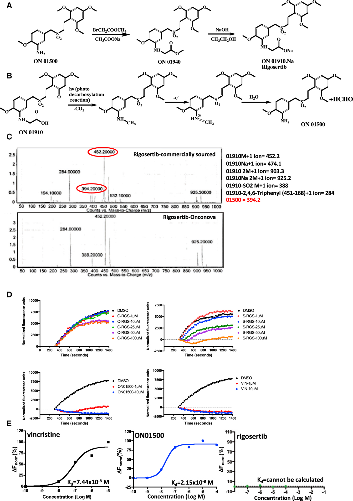
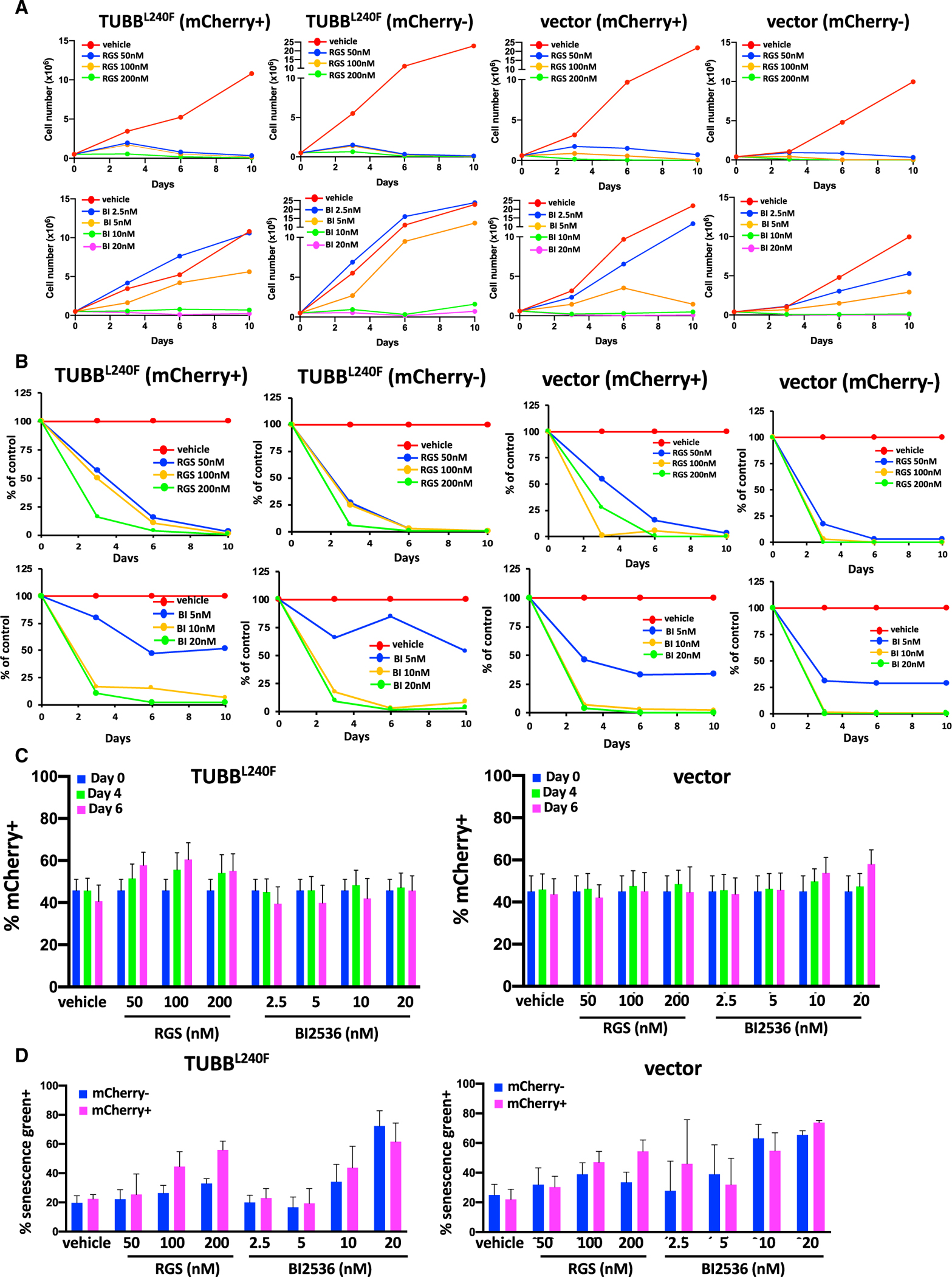
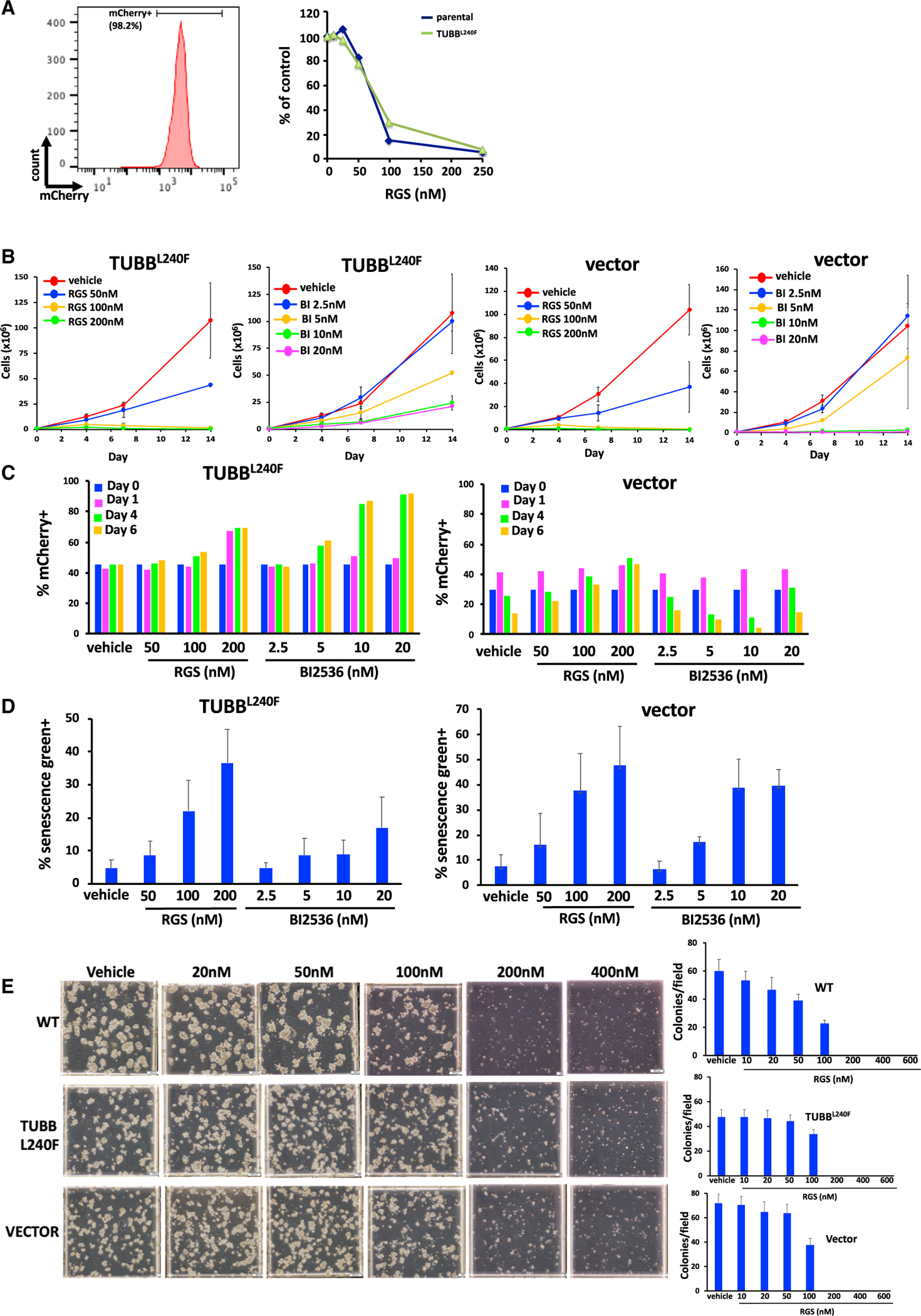
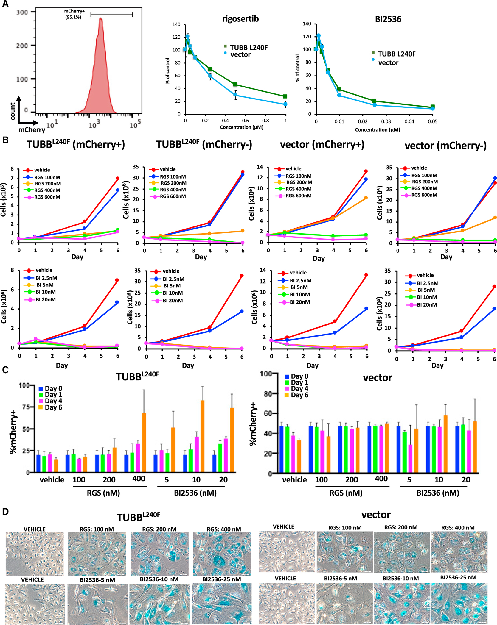
Rigosertib is a styryl benzyl sulfone that inhibits growth of tumor cells and acts as a RAS mimetic by binding to Ras binding domains of RAS effectors. A recent study attributed rigosertib's mechanism of action to microtubule binding. In that study, rigosertib was obtained from a commercial vendor. We compared the purity of clinical-grade and commercially sourced rigosertib and found that commercially sourced rigosertib contains approximately 5% ON01500, a potent inhibitor of tubulin polymerization. Clinical-grade rigosertib, which is free of this impurity, does not exhibit tubulin-binding activity. Cell lines expressing mutant β-tubulin have also been reported to be resistant to rigosertib. However, our study showed that these cells failed to proliferate in the presence of rigosertib at concentrations that are lethal to wild-type cells. Rigosertib induced a senescence-like phenotype in the small percentage of surviving cells, which could be incorrectly scored as resistant using short-term cultures.

Keywords: ON01500; ON01910; RAS; Ras binding domain; rigosertib; tubulin polymerization.


Conflict of interest statement

Declaration of Interests E.P.R. is an equity holder, board member, and paid consultant of Onconova Therapeutics. S.J.B. is a paid consultant of Onconova Therapeutics. M.V.R.R. and S.C.C. are stockholders and paid consultants of Onconova Therapeutics, Inc. M.V.R.R. and E.P.R. are named inventors on pending and/or issued patents filed by Temple University.

Figures

Figure 1.
Figure 1.. Purity and Tubulin-Binding Activities of Pharmaceutical-Grade and Commercial-Grade Rigosertib
(A) Synthetic scheme used for preparation of rigosertib (Reddy et al., 2011). (B) Photodegradation of rigosertib to ON01500. (C) LC-MS/MS analysis of rigosertib obtained from Onconova Therapeutics and Selleckchem. (D) Polymerization of tubulin in the presence of rigosertib from Onconova Therapeutics (O-RGS) and Selleckchem (S-RGS), ON01500 (Onconova Therapeutics), and vincristine (VIN). 25 μg of MAP-rich tubulin in general tubulin buffer was mixed with 1 mM guanosine triphosphate (GTP) and fluorescence reporter in the presence of vehicle (DMSO) or increasing concentrations of the indicated compound. Tubulin polymerization as a function of fluorescence was recorded over the indicated time at 37°C. (E) Microscale thermophoretic analysis of ON01500 and VIN with purified tubulin. Tubulin was labeled using the Monolith NT protein labeling kit RED-NHS according to the instructions of the manufacturer. Labeled protein was incubated with increasing concentrations of rigosertib, ON01500, or VIN for 30 min and subjected to MST.
Figure 2.
Figure 2.. 2Fo-Fc Electron Density Map (Gray) for Rigosertib Modeled in the Two αβ-Tubulin Heterodimers in the Asymmetric Unit of PDB: 5OV7
Density is contoured at 0.5σ (left), 0.8s (middle), and at 1σ (right).
Figure 3.
Figure 3.. Expression of Mutant β-Tubulin Does Not Confer a Significant Survival Advantage in K562 Cells Treated with Rigosertib
(A and B) Treatment with rigosertib induces a dose-dependent decrease in (A) proliferation and (B) viability of mCherry-positive TUBBL240F and WT K562 cells over time. K562 cells were infected with lentiviruses encoding β-tubulin L240F or an empty vector and combined with WT (mCherry−) cells at a final density of 1.0 × 10⁵ cells/mL. The cells were then treated with increasing concentrations of rigosertib, BI2536, or vehicle (DMSO) and harvested on days 1, 4, and 6. The percentages of viable (DAPI−) mCherry+ cells were determined using flow cytometric analysis. (C) Percentage of mCherry+ K562 cells expressing TUBBL240F as a function of time. K562 cells were infected with lentiviruses encoding mutant L240F β-tubulin or a vector control for 24 h. The percentage of mCherry+ cells was determined by flow cytometry analysis 48 h post-infection, and the cells were combined with WT (mCherry−) cells at a final density of 1.0 × 10⁵ cells/mL. The cells were then treated with increasing concentrations of rigosertib, BI2536, or vehicle (DMSO). Cells were harvested on days 4 and 6, and the percentage of viable (DAPI−) mCherry+ cells was determined using flow cytometric analysis. Error bars represent mean ± SEM. (D) Treatment with rigosertib induces senescence in WT and TUBBL240F K562 cells. K562 cells were infected with lentiviruses as described in (A) and grown in the presence of increasing concentrations of rigosertib or BI2536 for a 7-day period. mCherry+ and mCherry− cells were isolated using fluorescence-activated cell sorting and fixed, and the level of β-galactosidase was determined by staining with CellEvent senescence green and subsequent flow cytometric analysis. Data are represented as mean ± SEM.
Figure 4.
Figure 4.. Expression of L240F Mutant β-Tubulin Does Not Confer Resistance to Rigosertib
K562 cells were infected with a lentivirus that encodes the L240F mutant form of β-tubulin along with the mCherry fluorescence marker, and stable cell lines were isolated using limiting dilution cloning. (A) Flow cytometric analysis of mCherry expression in a representative K562 clone showing that more than 95% of cells are mCherry+ (left panel). These cells were then treated with the indicated concentrations of rigosertib, and the percentage of viable cells was determined 96 h after plating. Expression of the L240F β-tubulin mutant confers little or no resistance to rigosertib (right panel). (B) Growth of L240F β-tubulin-expressing K562 cells in the presence of increasing concentrations of rigosertib or BI2536, showing that rigosertib inhibits proliferation of K562 cells that express TUBBL240F. Analysis of one representative clone of each cell line is shown. Error bars represent mean ± SEM. (C) K562 cells expressing TUBBL240F were combined with WT cells and seeded at a density of 1 × 10⁵ cells/mL. The cells were treated with the indicated concentrations of rigosertib or BI2536 over a 6-day period, and the percentage of mCherry+ cells was determined by flow cytometry. The percentage of mCherry+ cells also increases in the presence of BI2536, indicating that this is not a rigosertib-specific phenomenon. (D) Treatment of K562 cells expressing TUBBL240F or a control vector with rigosertib or BI2536 induces senescence. Cells were treated with the indicated concentrations of rigosertib or BI2536 for a 7-day period, and the level of β-galactosidase activity in viable cells was measured by flow cytometric analysis using CellEvent senescence green. Error bars represent mean ± SEM. (E) 3D growth of L240F β-tubulin-expressing and control K562 cell lines in the presence of increasing concentrations of rigosertib, showing that rigosertib inhibits their proliferation in methylcellulose. Representative images (left panel) and average quantitation of 10 fields per plate in duplicate (right panels) are shown. Data are represented as mean ± SEM.
Figure 5.
Figure 5.. Expression of Mutant β-Tubulin Does Not Induce Resistance to Rigosertib in A549 Cells
A549 cells were infected with a lentivirus that encodes the L240F mutant form of β-tubulin along with the mCherry fluorescence marker. (A) Flow cytometry analysis of mCherry expression in representative A549 clones that are more than 95% mCherry+ (left panel). The cells were then treated with the indicated concentrations of rigosertib or BI2536, and their viability was determined 96 h post-plating (right panel). Error bars represent mean ± SEM. (B) A549 cells expressing TUBBL240F or a vector control were combined with WT A549 cells, grown in the presence of increasing concentrations of rigosertib or BI2536, and the growth of mCherry+ and mCherry− cells was measured over a 6-day period. As observed with K562 cells, rigosertib inhibits proliferation and survival of A549 cells that express TUBBL240F. Error bars represent mean ± SEM. (D) Prolonged treatment with rigosertib and BI2536 induces senescence in A549 cells. A549 cells expressing TUBB L240F or control cells were seeded at a density of 2 × 10⁵ cells per well in a 6-well dish. The cells were then treated with the indicated concentrations of rigosertib or BI2536 24 h post-plating. The medium was changed weekly. After 16 days in culture, the cells were washed with PBS, fixed, and stained overnight with X-gal (0.1 mg/mL) at 37°C. Data are represented as mean ± SEM.

References

    1. Agoni L, Basu I, Gupta S, Alfieri A, Gambino A, Goldberg GL, Reddy EP, and Guha C (2014). Rigosertib is a more effective radiosensitizer than cisplatin in concurrent chemoradiation treatment of cervical carcinoma, in vitro and in vivo. Int. J. Radiat. Oncol. Biol. Phys. 88, 1180–1187. - PMC - PubMed
    1. Athuluri-Divakar SK, Vasquez-Del Carpio R, Dutta K, Baker SJ, Cosenza SC, Basu I, Gupta YK, Reddy MV, Ueno L, Hart JR, et al. (2016). A Small Molecule RAS-Mimetic Disrupts RAS Association with Effector Proteins to Block Signaling. Cell 165, 643–655. - PMC - PubMed
    1. Driscoll DL, Chakravarty A, Bowman D, Shinde V, Lasky K, Shi J, Vos T, Stringer B, Amidon B, D’Amore N, and Hyer ML (2014). Plk1 inhibition causes post-mitotic DNA damage and senescence in a range of human tumor cell lines. PLoS ONE 9, e111060. - PMC - PubMed
    1. Jimeno A, Chan A, Cusatis G, Zhang X, Wheelhouse J, Solomon A, Chan F, Zhao M, Cosenza SC, Ramana Reddy MV, et al. (2009). Evaluation of the novel mitotic modulator ON 01910.Na in pancreatic cancer and preclinical development of an ex vivo predictive assay. Oncogene 28, 610–618. - PMC - PubMed
    1. Jost M, Chen Y, Gilbert LA, Horlbeck MA, Krenning L, Menchon G, Rai A, Cho MY, Stern JJ, Prota AE, et al. (2017). Combined CRISPRi/a-Based Chemical Genetic Screens Reveal that Rigosertib Is a Microtubule-Destabilizing Agent. Mol. Cell 68, 210–223.e6. - PMC - PubMed

Publication types