. 2020 Aug 17;30(16):3200-3211.e8.
doi: 10.1016/j.cub.2020.05.077. Epub 2020 Jul 2.

Input Connectivity Reveals Additional Heterogeneity of Dopaminergic Reinforcement in Drosophila

Affiliations

Input Connectivity Reveals Additional Heterogeneity of Dopaminergic Reinforcement in Drosophila

Nils Otto et al. Curr Biol. .

Abstract

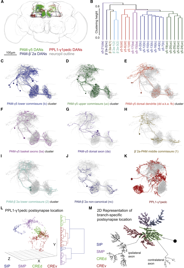
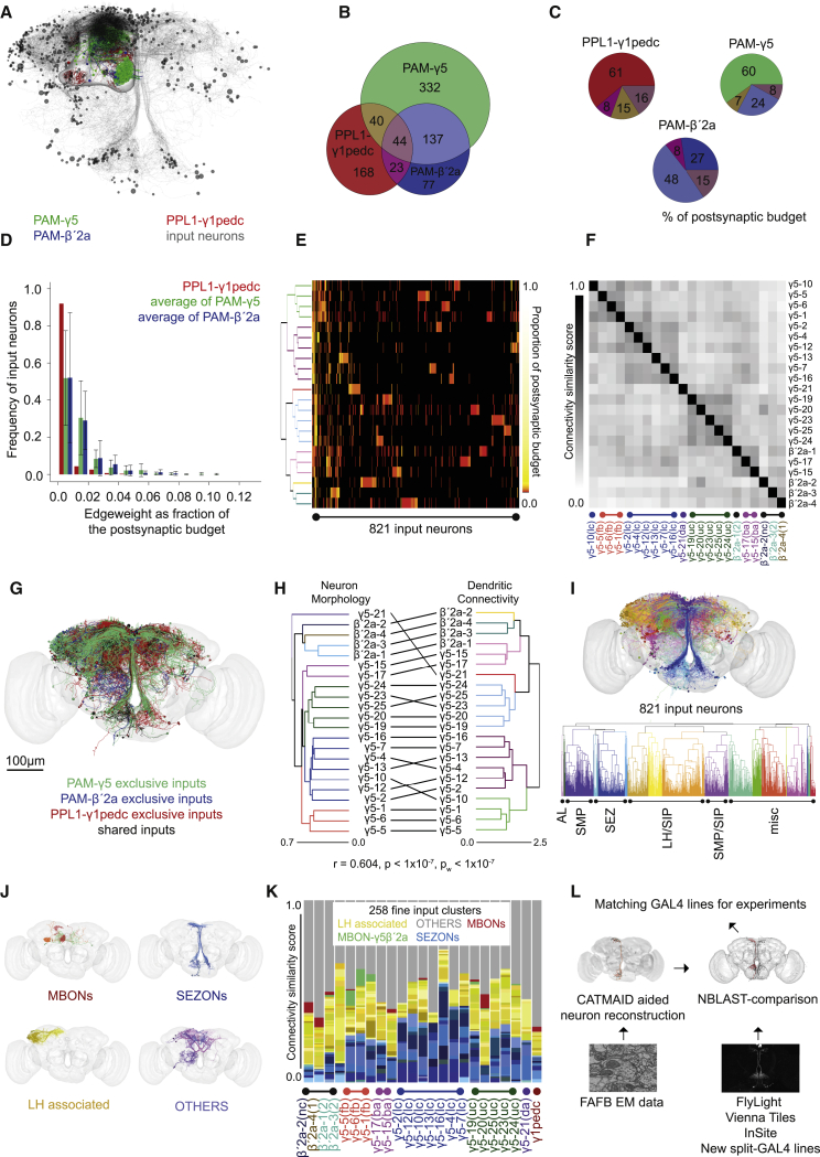
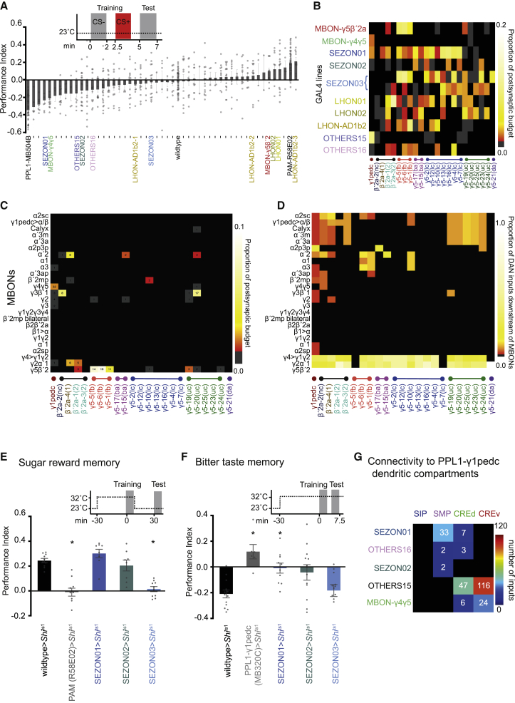
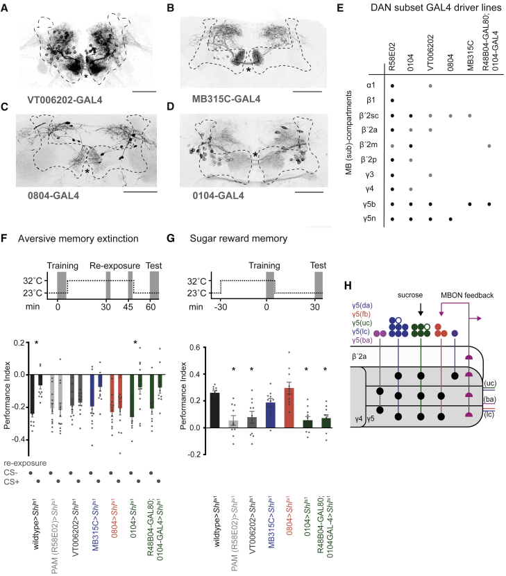
Different types of Drosophila dopaminergic neurons (DANs) reinforce memories of unique valence and provide state-dependent motivational control [1]. Prior studies suggest that the compartment architecture of the mushroom body (MB) is the relevant resolution for distinct DAN functions [2, 3]. Here we used a recent electron microscope volume of the fly brain [4] to reconstruct the fine anatomy of individual DANs within three MB compartments. We find the 20 DANs of the γ5 compartment, at least some of which provide reward teaching signals, can be clustered into 5 anatomical subtypes that innervate different regions within γ5. Reconstructing 821 upstream neurons reveals input selectivity, supporting the functional relevance of DAN sub-classification. Only one PAM-γ5 DAN subtype γ5(fb) receives direct recurrent feedback from γ5β'2a mushroom body output neurons (MBONs) and behavioral experiments distinguish a role for these DANs in memory revaluation from those reinforcing sugar memory. Other DAN subtypes receive major, and potentially reinforcing, inputs from putative gustatory interneurons or lateral horn neurons, which can also relay indirect feedback from MBONs. We similarly reconstructed the single aversively reinforcing PPL1-γ1pedc DAN. The γ1pedc DAN inputs mostly differ from those of γ5 DANs and they cluster onto distinct dendritic branches, presumably separating its established roles in aversive reinforcement and appetitive motivation [5, 6]. Tracing also identified neurons that provide broad input to γ5, β'2a, and γ1pedc DANs, suggesting that distributed DAN populations can be coordinately regulated. These connectomic and behavioral analyses therefore reveal further complexity of dopaminergic reinforcement circuits between and within MB compartments.

Keywords: Drosophila; connectomics; dopamine; extinction; learning; memory; mushroom body; reward.


Conflict of interest statement

Declaration of Interests The authors declare no competing interests.

Figures

Figure 1
Figure 1
Nanoscale Morphology of PAM-γ5, PAM-β′2a, and PPL1-γ1pedc DANs Reveals New Anatomical Subtypes and Features (A) Representation of all DANs reconstructed in this study. Twenty PAM-γ5 DANs on the fly’s right and 9 on the left, 8 PAM-β′2a DANs on the right, and both left and right PPL1-γ1pedc DANs. The MB and overall brain are outlined. Neuropil reference, Figure S1A. (B) Dendrogram showing hierarchical clustering of PAM DANs by morphology with 5 PAM-γ5 DAN and 3 β′2a DAN clusters. (C–K) Projection views of clustered reconstructed DANs. The morphology of the other traced PAM-γ5, PAM-β′2a DANs are shown in overlap (gray), and the MB neuropil (compare to A) is indicated by a dashed outline. (C) The 7 DANs of the PAM-γ5(lc) cluster (blue). (D) Five DANs of the PAM-γ5(uc) cluster (green). (E) Three DANs of the PAM-γ5(dd) cluster (scarlet). Only these neurons receive feedback from MBON-γ5β′2a (Figure 2K) and thus are renamed PAM-γ5(fb). (F) The 2 DANs of the PAM-γ5(ba) cluster (lilac). These DANs occupy the middle commissure. (G) The single PAM-γ5(da) DAN (purple). (H) A PAM-β′2a(1) DAN (ochre), which also occupies the middle commissure. (I) Two PAM-β′2a(2) DANs (turquoise). (J) A non-canonical PAM-β′2(nc) DAN (navy). (K) A PPL1-γ1pedc DAN (maroon). (L) Clustering of PPL1-γ1pedc DAN postsynapses in 3D space generates 4 distinct groups localized in the SIP (superior intermediate protocerebrum), SMP, and both a dorsal and ventral portion of the CRE (CREd and CREv). The optimum number of clusters was determined by the silhouette method; see Figure S1J. (M) Correlation of postsynapse clusters with PPL1-γ1pedc dendritic branches shown on a 2D dendrogram presented in the graphviz neato layout. See also Figure S1 and Videos S1, S2, and S4.
Figure 2
Figure 2
Input Specificity to Dopaminergic Neurons Matches Anatomical Subtypes (A) Representation of all 821 input neurons to PAM-γ5, PAM-β′2a, and PPL1-γ1pedc DANs identified in this study. Cell bodies (black spheres) and processes (gray). DANs and the MB outline shown for reference (compare to Figure 1A). (B) Venn diagram of unique and common input neurons to the analyzed DANs. PPL1-γ1pedc receives largely different input to PAM-γ5 and PAM-β′2a DANs. PAM-γ5 and PAM-β′2a DANs have many common inputs and some are also shared by PPL1-γ1pedc. (C) Pie charts showing percentage of postsynaptic budget occupied by shared and unique input neurons to PAM-γ5, PAM-β′2a, and PPL1-γ1pedc DANs. Percentage of shared inputs across all three groups is 16%, 15%, and 8% for PPL1-γ1pedc, PAM-β′2a, and PAM-γ5 DANs, respectively. (D) Bar chart showing DANs have many inputs with very low edge weight and each representing a small fraction of their overall postsynaptic budget. Inputs contributing more of the postsynaptic budget (to the right of the graph) are more abundant for PAM-γ5 and PAM-β′2a DANs; PPL1-γ1pedc distribution is strongly left shifted (bars show mean ± SD). (E) DANs can be clustered by input connectivity (rows correspond to F). Heatmap shows every DAN has a group of unique input neurons represented by unique blocks in each row. Clustering of DANs mostly depends on lesser number of shared inputs compressed to the left edge of the heatmap. (F) A matrix where DANs are grouped by the similarity of their input connectivity has clear structure, i.e., significantly more organized than random connectivity (comparison to null model, p < 0.0001; see Methods S1). (G) Representation of traced input neurons labeled using the unique and common input anatomy determined in (B) (see also Figures S2A–S2D). (H) Tanglegram comparing DAN clustering by morphology (from Figure 1B) and clustering by input connectivity (left of E). Connectivity and morphology are not significantly independent of each other (Pearson’s correlation between the corresponding distance matrices, r = 0.604; Mantel test, p < 10−7; pw < 10−7 within only γ5 or β′2a group). (I) DAN input neurons clustered by morphology. Dendrogram below shows single neurons allocated to 20 major coarse clusters based on soma position and primary neurite tract. Approximate neuropil of origin is indicated: antennal lobe (AL), SMP, SEZ, LH/SIP, and SMP/SIP are marked. Many neurons originate from less explored neuropils (misc). (J) Fine clusters of exemplary neurons for the MBON, LHON, SEZON, and OTHERS classes of DAN inputs. (K) Bar plot showing respective number of MBON, LHON, SEZON, and OTHERS inputs to individual DANs, ordered according to cluster identity (Figure 1). In general, PAM-γ5 DANs receive about 35% of their input from SEZONs, and about 20% from LHONs. Only the 3 PAM-γ5(fb) DANs receive significant direct input from MBON-γ5β′2a (green segments). PAM-β′2a DANs receive about 15% from SEZONs and 35% from LHONs. One PAM-β′2a DAN also receives minor direct input from MBON-γ5β′2a. PPL1-γ1pedc DANs receive roughly equal LHON and SEZON input. (L) NBLAST compares CATMAID generated neuronal skeletons from FAFB to neurons labeled in confocal images of GAL4 expression patterns. See also Figure S2, Methods S1, and Video S3.
Figure 3
Figure 3
Functional Analyses of DAN Input Neurons (A) Forty-nine GAL4 driver lines with identified DAN input neurons were used to drive UAS-CsChrimson and screened for memory implantation by pairing neuronal activation with odor exposure. Flies were starved 18–26 h prior to training and tested for immediate memory performance. Lines emphasized in this study (mean ± SEM; individual data points are displayed as dots, either P.I. > 0.1 or P.I. < −0.1, or connecting from a neuropil of prior interest) are labeled (Figure S3A, fully labeled version). (B) Connectivity matrix between DANs ordered according to morphological cluster identity and neurons labeled in 10 GAL4 lines, corresponding to 11 input clusters (MBON-γ5β′2a and MBON-γ4γ5, SEZON01-03, LHON01-02, LHON-AD1b2, and OTHERS15-16). Valence of memory formed is reflected by input connectivity. (C) Direct MBON-DAN connectivity matrix. We identified several MBONs to provide input to specific DANs (note: we traced all inputs to 7 PAM-γ5, and 2 β′2a DANs with extensive review; Figure S2; Methods S1). Numbers indicate total synapse counts between MBONs and DANs. (D) Adding other traced DAN input neurons creates potential for indirect connectivity between some MBONs and specific subsets of DANs. Indirect connectivity matrix showing the number of DAN input neurons that are downstream of MBONs with at least 3 synapses between each. Columns are normalized by their sum. (E) Olfactory learning with sucrose reinforcement. Schematic: experimental timeline and temperature shift protocol. Blocking neuron output during training abolished 30 min appetitive memory specifically in SEZON03-GAL4; UAS-Shits1 and R58E02-GAL4; UAS-Shits1 flies (mean ± SEM, p < 0.0241 and 0.0089, respectively; one-way ANOVA, with Dunnett’s post hoc test, n = 10). (F) Olfactory learning with bitter (DEET) reinforcement. Schematic: experimental timeline and temperature shift protocol. Blocking neuron output during training impaired immediate aversive memory in MB320C-, SEZON01-, and SEZON02-GAL4; UAS-Shits1, but not in SEZON03; UAS-Shits1 flies (mean ± SEM, p < 0.0009, 0.0344, and 0.0170, respectively; one-way ANOVA with Dunnett’s post hoc test, n = 12). (G) Connectivity matrix to specific branches of the PPL1-γ1pedc dendrite reveals classes of input neurons have branch specificity. See also Figure S3, Data S1, and Video S3.
Figure 4
Figure 4
Aversive Memory Extinction and Sugar Learning Require Different Subsets of PAM-γ5 DANs (A) Brain from a VT006202-GAL4; UAS-GFP fly labels all 20 PAM-γ5 DANs and possibly some other PAM DANs (black). Three commissures are visible (asterisk). Brain co-stained with nc82 antibody (MB is outlined). Scale bar, 20 μm. (B) MB315C-GAL4; UAS-GFP specifically labels 8 PAM-γ5 DANs per hemisphere that cross the midline in the lower commissure. (C) 0804-GAL4 labels 5 PAM-γ5 DANs per hemisphere, previously “γ5 narrow,” that occupy the lower commissure. Scale bar, 20 μm. (D) 0104-GAL4; UAS-GFP labels PAM-γ5 DANs, previously named “γ5 broad,” that cross the midline in the upper and middle commissures. 0104 also labels some other PAM DANs [9]. (E) Table summarizing DAN expression in GAL4 lines used for behavior, modified from [9]. R48B04GAL80 refines the 0104-GAL4 expression [12], shown in the Data S1. (F) Aversive olfactory memory extinction. Schematic: experimental timeline and temperature shift protocol. Blocking neuron output during odor re-exposure impaired memory extinction in R58E02-, VT006202-, and 0804-GAL4; UAS-Shits1, but not in MB315C- or 0104-GAL4 ± GAL80; UAS-Shits1 flies. Bars show mean ± SEM. Asterisks denote p < 0.035 (wild-type) and p < 0.0176 (0104); one-way ANOVA with Tukey’s post hoc test, n = 10–12. (G) Olfactory learning with sucrose reinforcement. Schematic: experimental timeline and temperature shift protocol. Blocking neuron output during training impaired 30 min appetitive memory in R58E02-, VT006202-, and 0104-GAL4 ± GAL80; UAS-Shits1, but not MB315C- or 0804-GAL4; UAS-Shits1 flies. Bars show mean ± SEM. Asterisks denote p < 0.0003 (R58E02), p < 0.0004 (0104), p < 0.0018 (VT006202), and p < 0.0010 (R48B04GAL80; 0104-GAL4); one-way ANOVA with Dunnett’s post hoc test, n = 10. (H) Schematic of input pathways to PAM-γ5 and PAM-β′2a DANs and subtype innervation of the γ5 compartment. Colors correspond to clusters defined in Figure 1. Open circles represent 2 γ5 DANs that were identified but not further analyzed. See also Figure S4, Data S1, and Video S4.

Comment in

References

    1. Cognigni P., Felsenberg J., Waddell S. Do the right thing: neural network mechanisms of memory formation, expression and update in Drosophila. Curr. Opin. Neurobiol. 2018;49:51–58. - PMC - PubMed
    1. Aso Y., Hattori D., Yu Y., Johnston R.M., Iyer N.A., Ngo T.T., Dionne H., Abbott L.F., Axel R., Tanimoto H., Rubin G.M. The neuronal architecture of the mushroom body provides a logic for associative learning. eLife. 2014;3:e04577. - PMC - PubMed
    1. Aso Y., Sitaraman D., Ichinose T., Kaun K.R., Vogt K., Belliart-Guérin G., Plaçais P.Y., Robie A.A., Yamagata N., Schnaitmann C. Mushroom body output neurons encode valence and guide memory-based action selection in Drosophila. eLife. 2014;3:e04580. - PMC - PubMed
    1. Zheng Z., Lauritzen J.S., Perlman E., Robinson C.G., Nichols M., Milkie D., Torrens O., Price J., Fisher C.B., Sharifi N. A complete electron microscopy volume of the brain of adult Drosophila melanogaster. Cell. 2018;174:730–743.e22. - PMC - PubMed
    1. Krashes M.J., DasGupta S., Vreede A., White B., Armstrong J.D., Waddell S. A neural circuit mechanism integrating motivational state with memory expression in Drosophila. Cell. 2009;139:416–427. - PMC - PubMed

Publication types

LinkOut - more resources