Review
. 2020 Oct:94:102901.
doi: 10.1016/j.dnarep.2020.102901. Epub 2020 Jun 26.

Twenty years of t-loops: A case study for the importance of collaboration in molecular biology

Affiliations
Review

Twenty years of t-loops: A case study for the importance of collaboration in molecular biology

Ľubomír Tomáška et al. DNA Repair (Amst). 2020 Oct.

Abstract

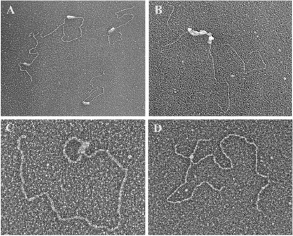
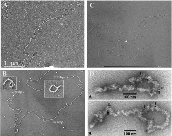
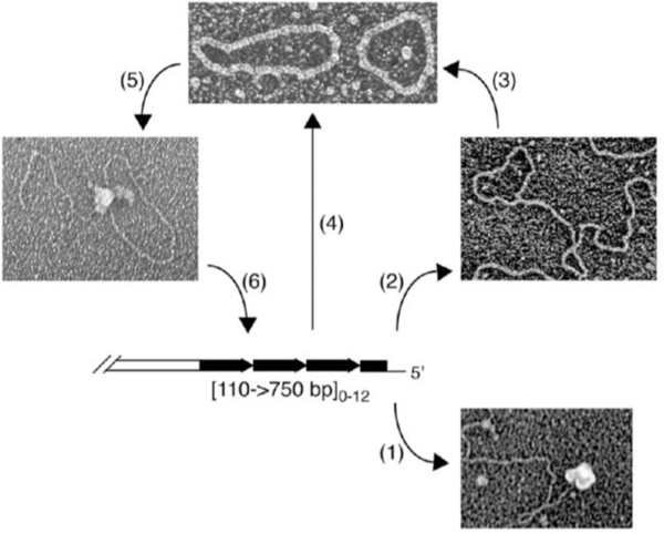
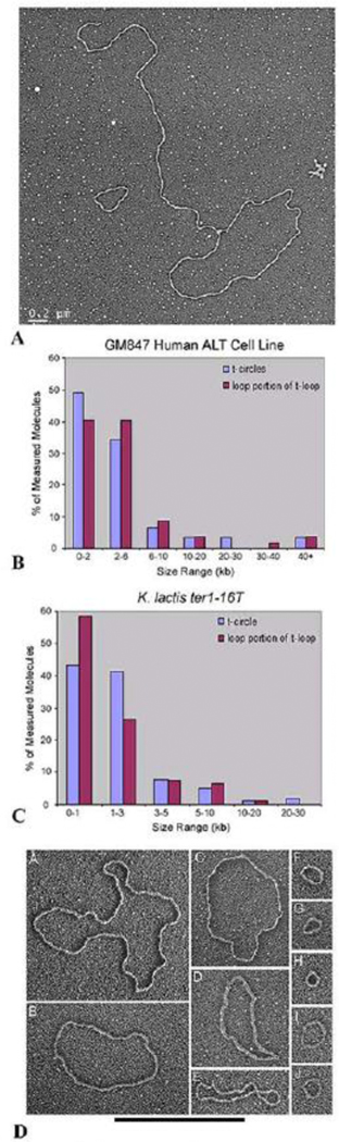
Collaborative studies open doors to breakthroughs otherwise unattainable by any one laboratory alone. Here we describe the initial collaboration between the Griffith and de Lange laboratories that led to thinking about the telomere as a DNA template for homologous recombination, the proposal of telomere looping, and the first electron micrographs of t-loops. This was followed by collaborations that revealed t-loops across eukaryotic phyla. The Griffith and Tomáška/Nosek collaboration revealed circular telomeric DNA (t-circles) derived from the linear mitochondrial chromosomes of nonconventional yeast, which spurred discovery of t-circles in ALT-positive human cells. Collaborative work between the Griffith and McEachern labs demonstrated t-loops and t-circles in a series of yeast species. The de Lange and Zhuang laboratories then applied super-resolution light microscopy to demonstrate a genetic role for TRF2 in loop formation. Recent work from the Griffith laboratory linked telomere transcription with t-loop formation, providing a new model of the t-loop junction. A recent collaboration between the Cesare and Gaus laboratories utilized super-resolution light microscopy to provide details about t-loops as protective elements, followed by the Boulton and Cesare laboratories showing how cell cycle regulation of TRF2 and RTEL enables t-loop opening and reformation to promote telomere replication. Twenty years after the discovery of t-loops, we reflect on the collective history of their research as a case study in collaborative molecular biology.

Keywords: DNA repair; Double strand breaks; R-loop; Super resolution microscopy; T-circle; T-loop; Telomeres.


Figures

Fig. 1.
Fig. 1.. TRF1 and TRF2 binding and sculpting DNA in vitro.
(A) Purified TRF1 binding to a linear ~3 kb plasmid DNA containing a single tract of 162 TTAGGG repeats roughly 1/3rd the distance from one end [30]. TRF1 fully coats the repeat tract. (B) TRF1 coating a 780 bp tract of TTRGGG repeats in the ~3 kb plasmid frequently results in a parallel synapsis of two DNAs at the repeat tract. Image from [30]. (C) TRF2 forms a t-loop on a model telomere DNA. The DNA backbone is ~3 kb and contains a 576 bp TTAGGG tract at one end including a 3’ ss G-rich overhang. The t-loop junction is coated with TRF2. (D) In this experiment from [39] a 3.5 kb pRST5 plasmid containing a replication fork within a 585 bp block of TTAGGG repeats was allowed to generate a chicken foot structure via slippage of the replication fork within the telomeric repeat tract. Addition of TRF2 revealed a strong preference for its binding at the Holliday junction on the chicken foot structure. Electron micrographs shown in reverse contrast, samples were directly mounted onto thin carbon foils, rotary shadow-cast with tungsten in a high vacuum and examined at 40 kV in a transmission electron microscope. See papers for details.
Fig. 2.
Fig. 2.. Examples of t-loop from across the phyla.
(A). T-loop isolated from HeLa 1.2.11 cells using the methods described in [37]. (B) Mini-chromosome from Trypanosoma brucei showing t-loops at both ends. The mini-chromosomes contain long TTAGGG tracts at each end. Image from [44]. (C) t-loop isolated from shoots and roots of common garden peas. Image from [45]. Small circular DNA is a 3 kb plasmid. Bar is equivalent to 10 kb in length. (D) Figure from [47] showing t-loops from chicken erythrocyte cells isolated as chromatin and stained with gold tagged beads carrying antibodies to TRF1.
Fig. 3.
Fig. 3.. T-loops and t-circles observed at yeast mitochondrial and nuclear telomeres.
(A) Several yeast species contain linear mitochondrial genomes with various types of organization of telomeres [116]. Here, mitochondrial telomeres are composed of an array of tandem repeats whose sequence and length is species-specific and a long (~100 nt) 5’ single-stranded overhang [51]. In C. parapsilosis this overhang can be protected by (1) mitochondrial telomere binding protein [–54], or (2) form a protective t-loop [58]. (3) T-loops may be resolved into t-circles that can also arise by (4) recombination between tandem repeats [55]. Once formed, (5) t-circles can undergo rolling-circle replication [57] generating a long array of telomeric repeats that (6) can integrate back into the main genome [59]
Fig. 4.
Fig. 4.. T-circles from human ALT and K. lactis cells.
(A) Micrograph of a t-loop and t-circle isolated from GM847 ALT cells following psoralen/UV crosslinking as in [37]. (B) Distribution of circle sizes measured from the circular portions of t-loops isolated from GM847 cells (red) or t-circles (blue) from the same cells. Data from [60]. (C) Distribution of circle sizes measured from the circular portions of t-loops isolated from K lactis cells (red) or t-circles (blue) from the same cells. Data from Cesare et al (2008) [62]. (D). Panel of examples of t-circles isolated from K. lactis (ter1–16T) cells. Image from [62].
Fig. 5.
Fig. 5.. T-loops in yeast: examples from in vitro and in vivo experiments.
(A) t-loops formed by purified Taz1 protein of S. pombe on a model telomere template containing a block of 518 bp of the S. pombe consensus sequence (5’-GGTTACA-3’) located at one end. Taz1 forms large donut-like oligomers which bind the telomeric repeats as described in [72]. Model DNAs measure 2.9 kb in length. Following end binding, the DNA appears to thread through the hole in the donut (inset panel A) followed by the donut sliding along the DNA to form a loop which can exceed the length of the telomeric tract. (B) Purified Tay1 protein from Y. lipolytica binds along a 810 bp tract of 5’-GGGTTAGTCA-3’ repeats at one end of a linear ~3 kb model telomere DNA. Tay1 coats the repeat tract in a mode similar to TRF1 binding, but also generates looped structures similar to the looping induced by TRF2. Image from [77]. (C) T-loop DNA isolated from ter1-16T mutant K. lactis cells containing long telomeres as described in [62]. Bar in C is equivalent to 3 kb.
Fig. 6.
Fig. 6.
Visualizing t-loops using light microscope STORM imaging. Montage of t-loop molecules imaged by STORM. From[38]. Courtesy of Ylli Doksani, IFROM, Milan, Italy.
Fig. 7.
Fig. 7.. Transcription drives t-loop formation.
(A) As described in [100] the ~3 kb plasmid pRST5 containing a 576 bp block of TTAGGG repeats at one end and immediately preceded by a T7 RNA polymerase promoter was transcribed in vitro followed by deproteinization and preparation for EM. Fields of DNAs showed up to 60% of the DNAs with a small t-loop at the end containing the telomeric repeats. (B) In [100] a rolling circle replication scheme was used to generate double stranded telomeric DNA exceeding 10 kb in length and containing a T7 promoter at one end. Its transcription in vitro frequently generated t-loop structures with dimensions very similar to ones isolated from human cells. (C) Model of how t-loops may form due to transcription and R-loop formation [100].

Comment in

References

    1. Muller HJ, The remaking of chromosomes. Collecting net, 1938. 13: p. 181–198.
    1. McClintock B, The Stability of Broken Ends of Chromosomes in Zea Mays. Genetics, 1941. 26(2): p. 234–82. - PMC - PubMed
    1. de Lange T, Shelterin-Mediated Telomere Protection. Annu Rev Genet, 2018. 52: p. 223–247. - PubMed
    1. Olovnikov AM, A theory of marginotomy. The incomplete copying of template margin in enzymic synthesis of polynucleotides and biological significance of the phenomenon. J Theor Biol, 1973. 41(1): p. 181–90. - PubMed
    1. Watson JD, Origin of concatemeric T7 DNA. Nat New Biol, 1972. 239(94): p. 197–201. - PubMed

Publication types

MeSH terms