Review
. 2020 Jul 3;147(13):dev183020.
doi: 10.1242/dev.183020.

Mechanisms of heart valve development and disease

Affiliations
Review

Mechanisms of heart valve development and disease

Anna O'Donnell et al. Development. .

Abstract

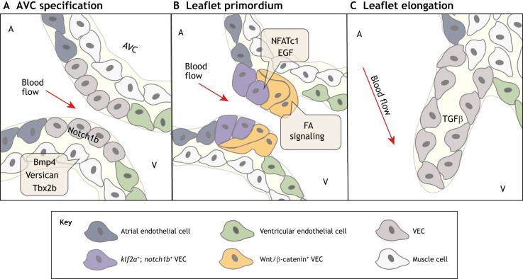
The valves of the heart are crucial for ensuring that blood flows in one direction from the heart, through the lungs and back to the rest of the body. Heart valve development is regulated by complex interactions between different cardiac cell types and is subject to blood flow-driven forces. Recent work has begun to elucidate the important roles of developmental pathways, valve cell heterogeneity and hemodynamics in determining the structure and function of developing valves. Furthermore, this work has revealed that many key genetic pathways involved in cardiac valve development are also implicated in diseased valves. Here, we review recent discoveries that have furthered our understanding of the molecular, cellular and mechanosensitive mechanisms of valve development, and highlight new insights into congenital and acquired valve disease.

Keywords: Congenital valve disease; Heart valve development; Hemodynamics.


Conflict of interest statement

Competing interestsThe authors declare no competing or financial interests.

Figures

Fig. 1.
Fig. 1.
Cardiac valves in the adult mammalian and the zebrafish heart. (A) The mammalian heart consists of four chambers – the left and right atria, and the left and right ventricles – separated by four valves. The AV valves, which include the mitral valve (green) and the tricuspid valve (dark blue), lie between the atria and the ventricles; the SL valves, which include the pulmonary valve (dark red) and the aortic valve (yellow), lie between the ventricles and the outflow tracts. (B) The zebrafish heart consists of two chambers – one atrium and one ventricle – and two valves. The atrioventricular valve (cyan), which is similar to the mammalian tricuspid and mitral valves, lies between the atrium and the ventricle; the bulboventricular valve (orange), which is similar to the mammalian aortic valve, lies between the ventricle and the bulbus arteriosus (purple). AV, atrioventricular; SL, semilunar.
Fig. 2.
Fig. 2.
The microarchitecture of adult cardiac valves. (A,B) The stratified structure of aortic (A) and mitral (B) cardiac valves ensures unidirectional blood flow. Valve leaflets are composed of three layers of stratified extracellular matrix, including an elastin-rich ventricularis layer in the SL valves or the atrialis layer in the AV valves (purple), a proteoglycan-rich spongiosa layer (light blue) and a collagen-rich fibrosa layer (yellow), interspersed with valve interstitial cells (VICs) and sheathed in a monolayer of valve endothelial cells (VECs). The direction of pulsatile blood flow in relation to valve leaflets is indicated (red arrows). The microarchitecture of an aortic valve leaflet ensures blood moves from the ventricle to the aorta. The microarchitecture of a mitral valve leaflet is supported by the chordae tendineae, which ensure blood flow from the atrium to the ventricle. AV, atrioventricular; SL, semilunar.
Fig. 3.
Fig. 3.
The molecular pathways regulating endocardial cushion formation, endothelial-to-mesenchymal transition (EndoMT) and valve primordia growth. (A) E9-10: endocardial cushion formation is initiated when myocardial Bmp2 promotes the expression of hyaluronan, Tbx2 and Tbx3 to stimulate cardiac jelly formation in endocardial swellings in the atrioventricular canal (AVC). (B) E10-12: myocardial BMP signaling, through endocardial Bmpr1a and endocardial Notch1, and Wnt/β-catenin, TGFβ and Hippo/Yap1 signaling promote EndoMT and the expression of the mesenchymal markers Twist, Msx1/2 and Snail. (C) E14-18: the distal proliferation of valve endothelial cells (VECs) and valve interstitial cells (VICs) stimulates the outgrowth of leaflet primordia. VEC proliferation is induced by Wnt/β-catenin and VEGF signaling. VIC proliferation is induced by BMP and FGF4 signaling, and is inhibited by EGF and Notch signaling.
Fig. 4.
Fig. 4.
Postnatal aortic valve endothelial and interstitial subpopulations. Postnatal aortic valves contain localized subpopulations of valve endothelial cells (VECs), valve interstitial cells (VICs) and immune cells that exhibit distinct transcriptional profiles. These populations are present in regions of the valve exposed to unique combinations of mechanical forces. The typical VEC subpopulation (gray) can be found on both sides of the valve leaflet, while a VEC subpopulation expressing Prox1 (yellow), a lymphatic marker influenced by oscillatory shear stress, is found solely on the side away from laminar flow. A coaptation VEC population (orange) is found where the leaflets meet. A collagen-expressing subpopulation of VICs (pink) is found in the fibrosa layer and a GAG-expressing subpopulation of VICs (green) is in the hinge and tip of the leaflet. Similarly, macrophages are present in the hinge and tip regions of the valve leaflet, which are subject to high mechanical stress. GAG, glycosaminoglycan.
Fig. 5.
Fig. 5.
Molecular pathways regulating zebrafish valve development. (A) At ∼36-48 hpf, the restriction of bmp4, versican and tbx2 in the myocardium and notch1b in the endocardium initiates atrioventricular canal (AVC) specification between the atrium (A) and the ventricle (V). (B) At ∼50-60 hpf, endocardial cushion-like valve primordia then form through partial endothelial-to-mesenchymal transition (EndoMT), apparent by endocardial invagination and collective migration. klf2a- and notch1b-expressing valve endothelial cell (VEC) progenitors on the ventricular side of the AVC undergo EndoMT or invaginate under notch1b-expressing valve progenitors on the atrial side of the AVC. The NFATc1, Wnt, Notch, EGF and focal adhesion (FA) signaling pathways have also been implicated in the formation of valve primordia. (C) At ∼100 hpf-adult, valve primordia then elongate and thin in response to TGFβ signaling, and remain thin with little extracellular matrix until 28 dpf. The direction of pulsatile blood flow in relation to developing valve structures is indicated (red arrow). hpf, hours post fertilization.

References

    1. Aikawa E., Whittaker P., Farber M., Mendelson K., Padera R. F., Aikawa M. and Schoen F. J. (2006). Human semilunar cardiac valve remodeling by activated cells from fetus to adult: implications for postnatal adaptation, pathology, and tissue engineering. Circulation 113, 1344-1352. 10.1161/CIRCULATIONAHA.105.591768 - DOI - PubMed
    1. Aikawa E., Nahrendorf M., Sosnovik D., Lok V. M., Jaffer F. A., Aikawa M. and Weissleder R. (2007). Multimodality molecular imaging identifies proteolytic and osteogenic activities in early aortic valve disease. Circulation 115, 377-386. 10.1161/CIRCULATIONAHA.106.654913 - DOI - PubMed
    1. Alfieri O. and Vahanian A. (2017). The year in cardiology 2016: valvular heart disease. Eur. Heart J. 38, 628-633. 10.1093/eurheartj/ehw636 - DOI - PubMed
    1. Armstrong E. J. and Bischoff J. (2004). Heart valve development: endothelial cell signaling and differentiation. Circ. Res. 95, 459-470. 10.1161/01.RES.0000141146.95728.da - DOI - PMC - PubMed
    1. Aupperle H. and Disatian S. (2012). Pathology, protein expression and signaling in myxomatous mitral valve degeneration: comparison of dogs and humans. J. Vet. Cardiol. 14, 59-71. 10.1016/j.jvc.2012.01.005 - DOI - PubMed

Publication types

MeSH terms