. 2020 Jul 3;3(1):342.
doi: 10.1038/s42003-020-1072-4.

Salmonella-based platform for efficient delivery of functional binding proteins to the cytosol

Affiliations

Salmonella-based platform for efficient delivery of functional binding proteins to the cytosol

Antoine Chabloz et al. Commun Biol. .

Abstract

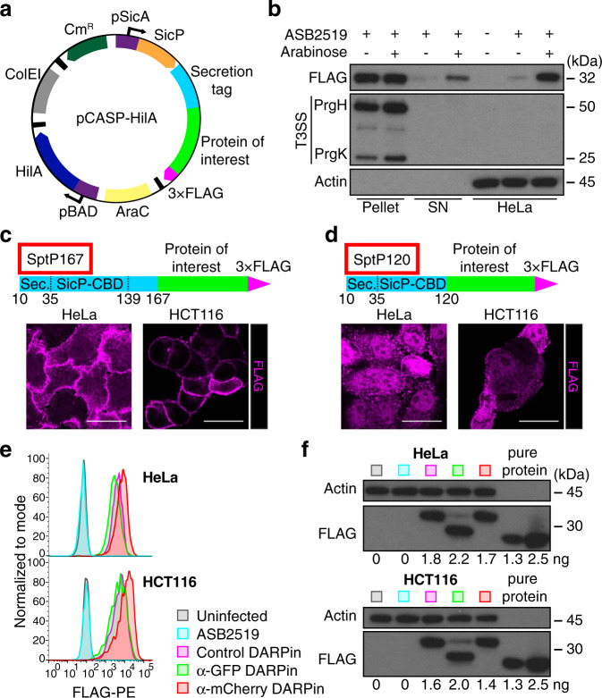
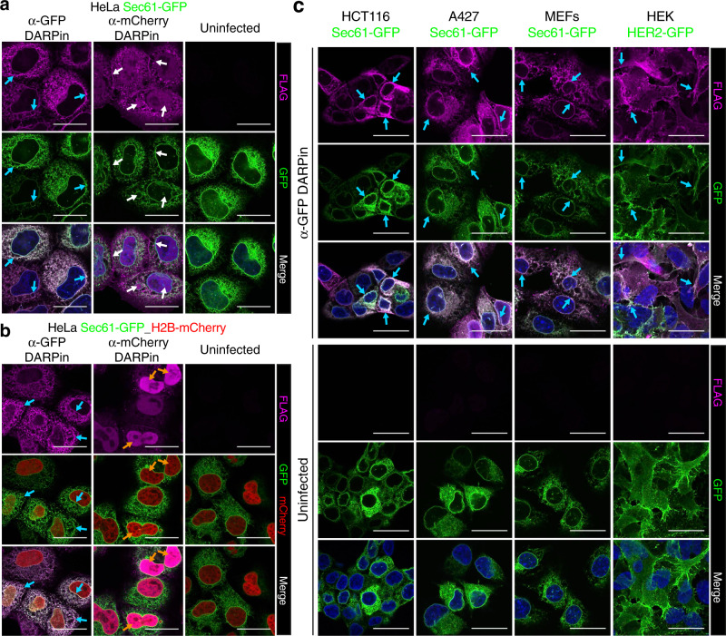
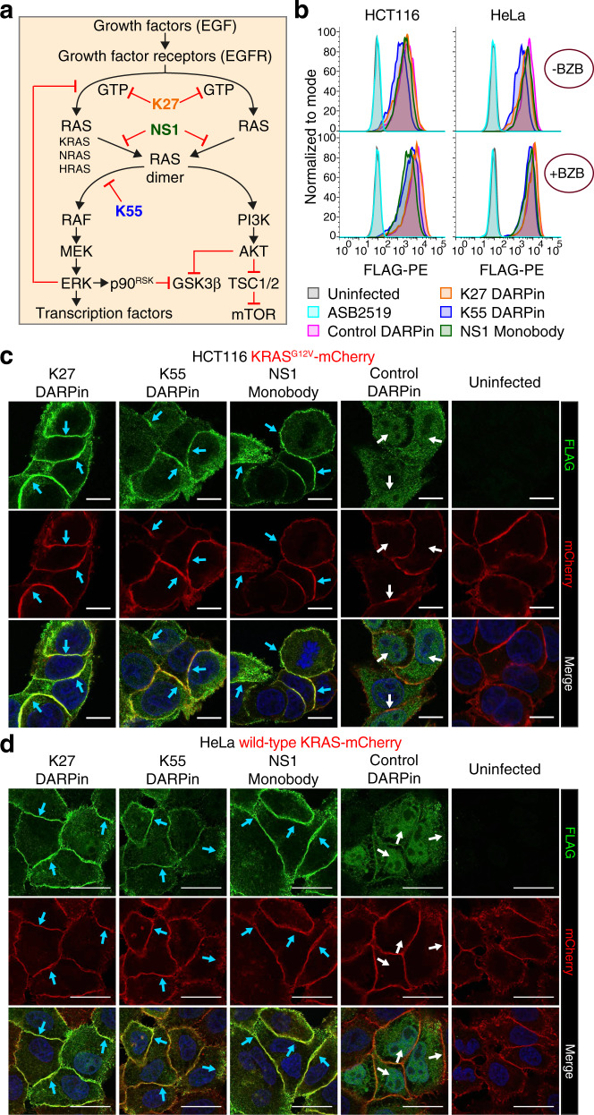
Protein-based affinity reagents (like antibodies or alternative binding scaffolds) offer wide-ranging applications for basic research and therapeutic approaches. However, whereas small chemical molecules efficiently reach intracellular targets, the delivery of macromolecules into the cytosol of cells remains a major challenge; thus cytosolic applications of protein-based reagents are rather limited. Some pathogenic bacteria have evolved a conserved type III secretion system (T3SS) which allows the delivery of effector proteins into eukaryotic cells. Here, we enhance the T3SS of an avirulent strain of Salmonella typhimurium to reproducibly deliver multiple classes of recombinant proteins into eukaryotic cells. The efficacy of the system is probed with both DARPins and monobodies to functionally inhibit the paradigmatic and largely undruggable RAS signaling pathway. Thus, we develop a bacterial secretion system for potent cytosolic delivery of therapeutic macromolecules.


Conflict of interest statement

The authors declare no competing interests.

Figures

Fig. 1
Fig. 1. Improvement of the T3SS into an efficient cytosolic delivery system.
a Schematic representation of the pCASP-HilA secretion system plasmid. The translation of the SptP specific chaperone SicP (in orange) and the fusion protein from c or d are coupled on a single mRNA and regulated by the Salmonella pSicA promoter. The main regulator of SPI-1 genes HilA is under the regulation of an arabinose-inducible promoter (pBAD). Black boxes represent transcriptional termination and purple boxes with arrows represent promoters. CmR: chloramphenicol resistance, ColEI: origin of replication, AraC: Arabinose operon regulatory protein. b Anti-FLAG tag western blot of bacterial pellets and supernatants (SN) after 4 h induction of Salmonella ASB2519 with or without arabinose in the last 2 h, and HeLa cells infected with those Salmonella for 1 h at a MOI of 100 in the presence or absence of arabinose. Anti-needle complex (T3SS) and anti-Actin blots serve as control for the presence of Salmonella and eukaryotic cells, respectively. c, d (Top) Schematic of the transferred SptP167 and SptP120 3 × FLAG-tagged fusion recombinant proteins. Secretion signal (Sec.), SicP-chaperone binding domain (SicP-CBD) and amino acid numbers that mark the beginning and end of each SptP domain are indicated. Representative anti-FLAG-immunostaining images of anti-GFP (α-GFP) FLAG-tagged DARPins transferred into HeLa and HCT116 cells using the SptP167 (c) or SptP120 (d) secretion tags (1 h infection at a MOI of 100). Scale bars represent 25 µm. e Flow cytometry analysis of 3 different FLAG-tagged SptP120-DARPins (E3_5 control, α-GFP 3G124 and α-mCherry 3m160 DARPins) transferred into HeLa and HCT116 cells following 1 h infection at a MOI of 100. Uninfected cells and Salmonella ASB2519 with an empty pCASP-HilA vector (ASB2519) served as negative controls. Experiments were repeated in three biological replicates showing comparable results. f Quantitative anti-FLAG tag western blot of HeLa and HCT116 cells from the same experiment as in e. Numbers below the blots show quantification of the respective transferred SptP120-DARPin fusion proteins, based on 1.3 and 2.5 ng of a purified 3×FLAG protein (pure protein - RuvC) loaded on the same blot. Uncropped blots can be found in Supplementary Fig. 6.
Fig. 2
Fig. 2. Delivery of functional DARPins into multiple cell types by Salmonella ASB2519.
a, b Representative anti-FLAG-immunostaining images (magenta, upper panels) of anti-GFP (α-GFP) and α-mCherry FLAG-tagged SptP120-DARPins transferred into HeLa cells (1 h infection at a MOI of 100). Experiments were performed in HeLa cells expressing (a) Sec61-GFP or (b) both Sec61-GFP and H2B-mCherry. GFP (green) and mCherry (red) expression and merged images are also shown. c Representative anti-FLAG-immunostainings of α-GFP FLAG-tagged SptP120-DARPin transferred into the indicated human and mouse cell types expressing Sec61-GFP and HER2-GFP. Experiments were repeated in three biological replicates showing comparable results. Uninfected samples served as negative controls. Scale bars represent 25 µm. Blue and orange arrows indicate colocalization in the cytoplasm and in nuclei, respectively. White arrows show that the anti-FLAG signal does not co-localize with GFP using the α-mCherry DARPin.
Fig. 3
Fig. 3. Synthetic RAS inhibitory proteins are efficiently delivered into cells and co-localize with overexpressed mutated or wild-type KRAS proteins.
a Simplified RAS pathway overview with the DARPins (K27 and K55) and monobody (NS1) used to block RAS activation (modified from www.cancer.gov). Black arrows indicate activation and red lines show inhibition. b Flow cytometry analysis of the FLAG-tagged SptP120-control DARPin and SptP120-anti-RAS inhibitors transferred into HCT116 and HeLa cells (1 h infection at a MOI of 100), in the absence (upper histograms) or presence (lower histograms) of the proteasome inhibitor bortezomib (BZB; 50 nM). Uninfected cells and cells infected with Salmonella ASB2519 with an empty pCASP-HilA vector (ASB2519) served as negative controls. Representative anti-FLAG-immunostainings (green) of FLAG-tagged SptP120-anti-RAS inhibitors transferred into HCT116 (c) and HeLa (d) cells expressing KRASG12V or wild-type KRAS-mCherry. All samples were treated with BZB followed by 10 min incubation in fresh medium without (c) or with (d) EGF (20 ng per mL) stimulation. Experiments were repeated in two biological replicates showing comparable results. Uninfected cells and SptP120-control DARPin served as negative controls. Scale bars are (c) 10 µm or (d) 25 µm. Blue arrows indicate colocalization. White arrows show that the anti-FLAG signal does not co-localize with mCherry using the control DARPin.
Fig. 4
Fig. 4. T3SS-delivered RAS inhibitors downregulate RAS signaling.
a Western blot analysis of uninfected HCT116 cells (Uninf) and HCT116 cells infected with Salmonella ASB2519 (1 h, MOI of 100) delivering a FLAG-tagged SptP120-control (Ctrl) DARPin and the SptP120-anti-RAS inhibitory DARPins K27 and K55 or the RAS blocking monobody NS1. Anti-FLAG detection served as the transfer control and total anti-ERK1/2, AKT and Actin as loading controls. Numbers below the pERK1/2 and pAKT blots detected with anti-pERK1/2 (Thr202/Tyr204) and anti-pAKT (Ser473) antibodies represent the quantified fraction relative to the SptP120-control DARPin (Ctrl), normalized to total ERK1/2 and AKT, respectively. Uncropped blots can be found in Supplementary Fig. 10. b Flow cytometric measurements of ERK1/2 phosphorylation in HCT116 cells upon FLAG-positive delivery of the indicated SptP120-anti-RAS binders. Data were analyzed 10 min post-infection in the presence of bortezomib (BZB; 50 nM). Pink lines indicate baseline ERK1/2 phosphorylation levels upon delivery of the SptP120-control DARPin. Relative median fluorescence intensities (MFI) of ERK1/2 (c) and GSK3β (d) phosphorylation compared to the SptP120-control DARPin (Ctrl) treated cells and using the same experimental setup as in b. e Flow cytometric analysis of ERK1/2 phosphorylation in HeLa cells upon FLAG-positive delivery of the indicated SptP120-anti-RAS binders and a control DARPin. Data were analyzed 10 min post-infection in the presence of bortezomib (BZB) without (empty) or with EGF (20 ng per mL) stimulation (filled). Relative median fluorescence intensities (MFI) of ERK1/2 (f) and GSK3β (g) phosphorylation compared to the SptP120-control DARPin (Ctrl) treated cells with EGF and using the same experimental setup as in e. Data represent the mean ± SEM of six (c, d) or three (f, g) biological replicates. Individual data points are shown. Statistical analysis was performed using a one-way ANOVA with Tukey’s multiple comparisons test (****P< 0.0001; ***P< 0.001; **P< 0.01; *P< 0.05; ns not significant; only relevant comparisons are indicated for (f) and (g)).

References

    1. Vivès E, Brodin P, Lebleu B. A truncated HIV-1 Tat protein basic domain rapidly translocates through the plasma membrane and accumulates in the cell nucleus. J. Biol. Chem. 1997;272:16010–16017. - PubMed
    1. Derossi D, Joliot AH, Chassaing G, Prochiantz A. The third helix of the antennapedia homeodornain translocates through biological membranes. J. Biol. Chem. 1994;296:10444–10450. - PubMed
    1. Deprey K, Becker L, Kritzer J, Plückthun A. Trapped! a critical evaluation of methods for measuring total cellular uptake versus cytosolic localization. Bioconjug. Chem. 2019;30:1006–1027. - PMC - PubMed
    1. Kamaly N, Xiao Z, Valencia PM, Radovic-Moreno AF, Farokhzad OC. Targeted polymeric therapeutic nanoparticles: design, development and clinical translation. Chem. Soc. Rev. 2012;41:2971–3010. - PMC - PubMed
    1. Smith SA, Selby LI, Johnston APR, Such GK. The endosomal escape of nanoparticles: toward more efficient cellular delivery. Bioconjug. Chem. 2019;30:263–272. - PubMed

Publication types

MeSH terms