. 2020 Oct:89:245-255.
doi: 10.1016/j.bbi.2020.06.028. Epub 2020 Jul 2.

Microglial depletion aggravates the severity of acute and chronic seizures in mice

Affiliations

Microglial depletion aggravates the severity of acute and chronic seizures in mice

Wenning Wu et al. Brain Behav Immun. 2020 Oct.

Abstract

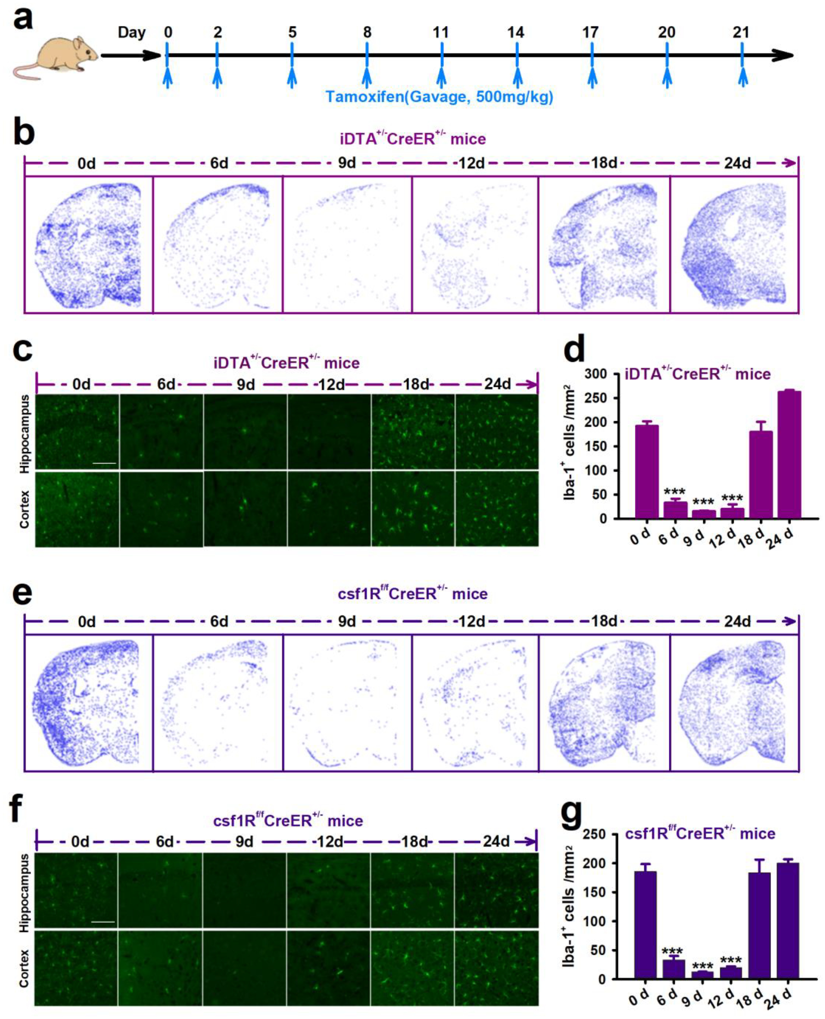
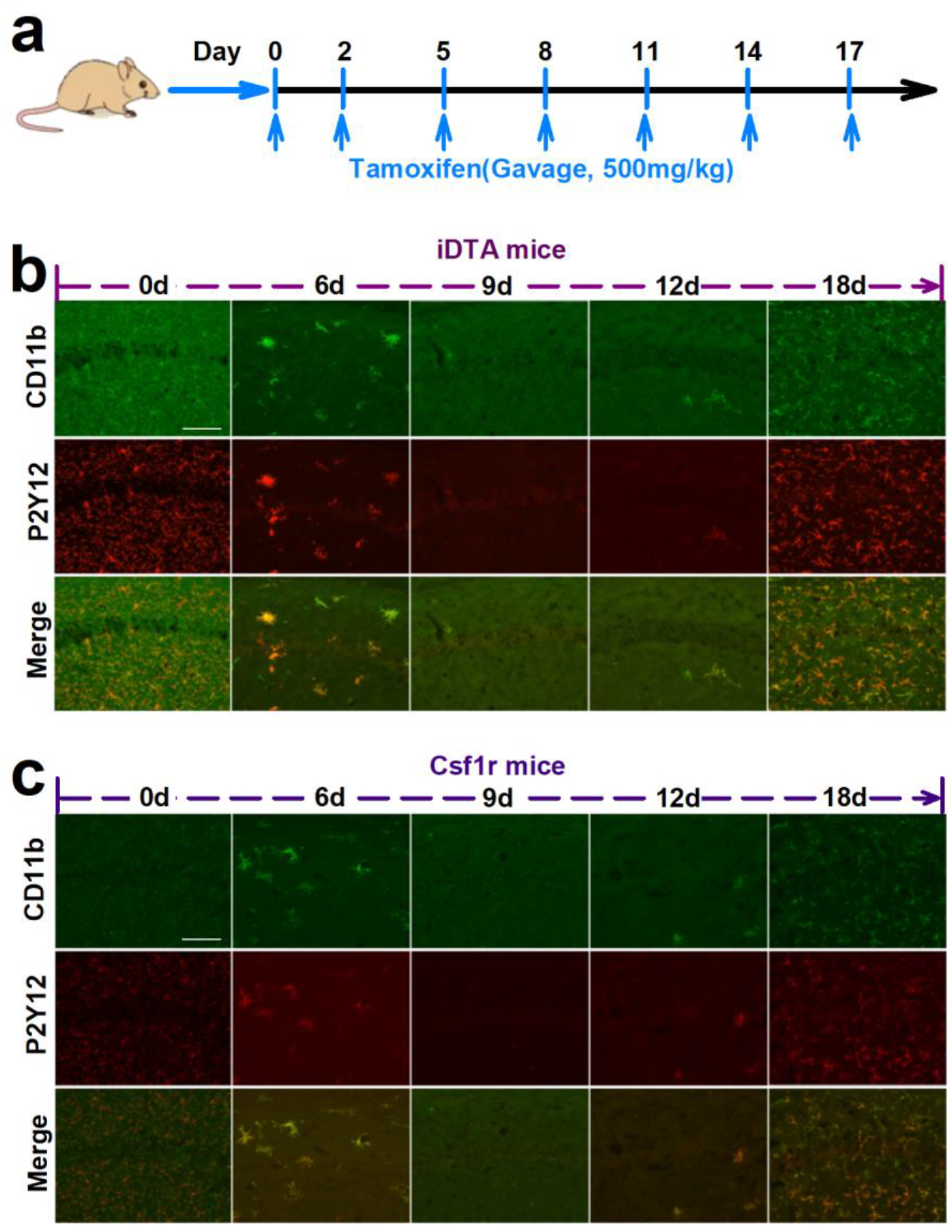
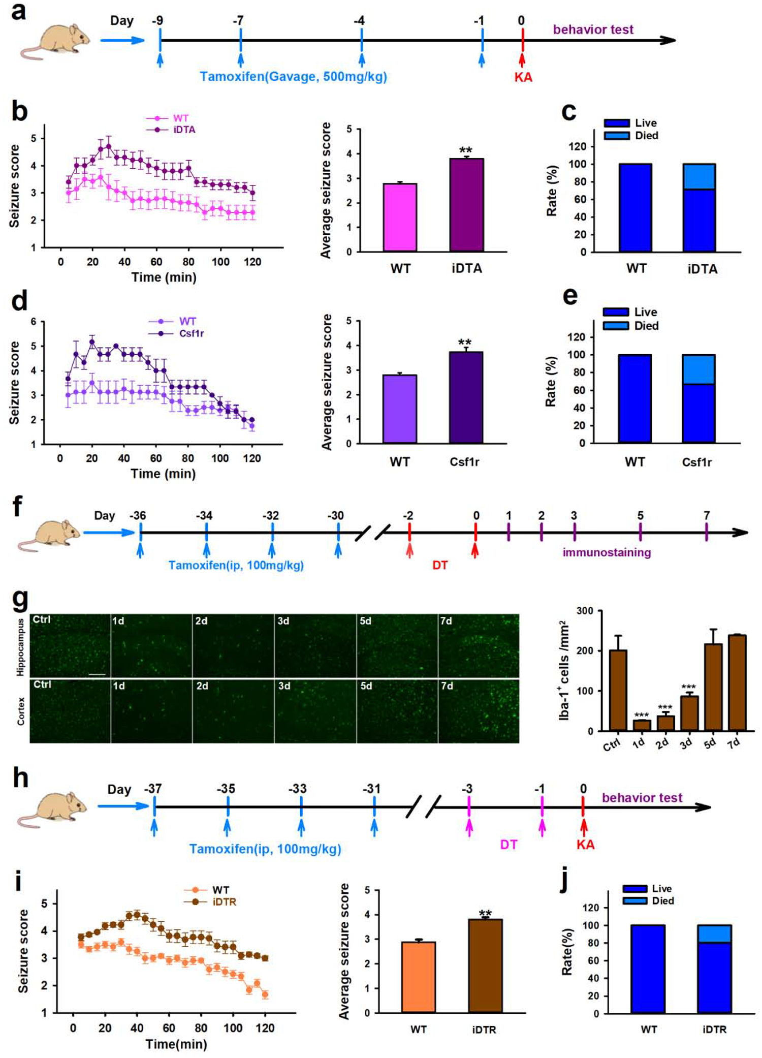
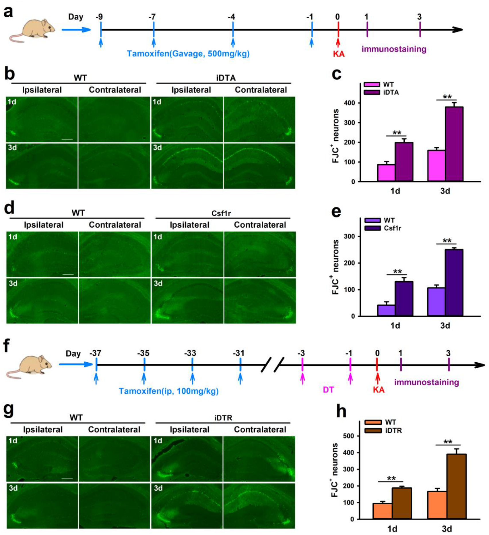
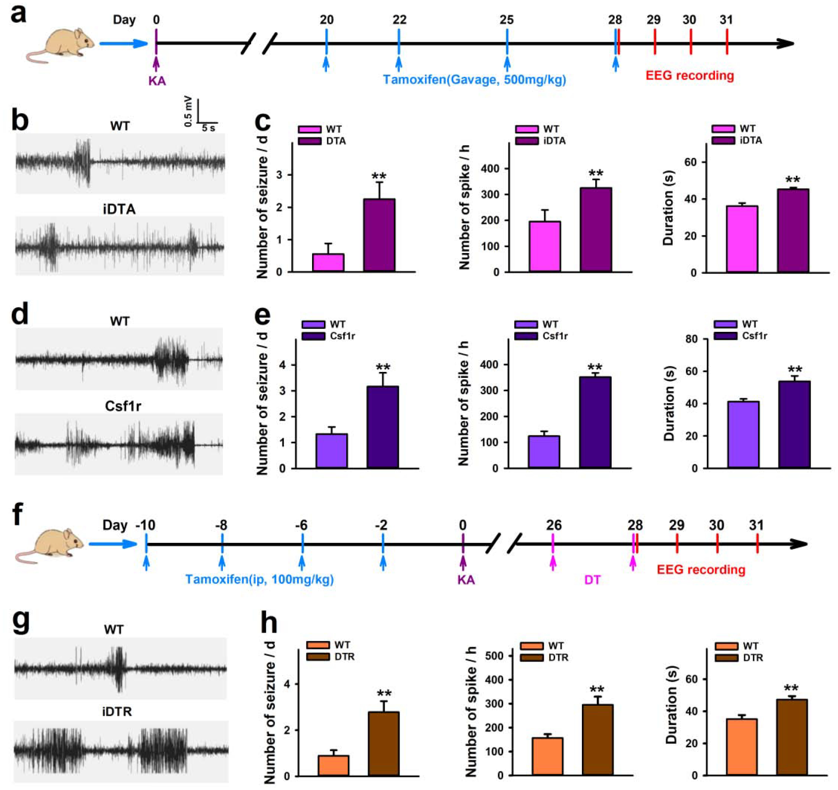
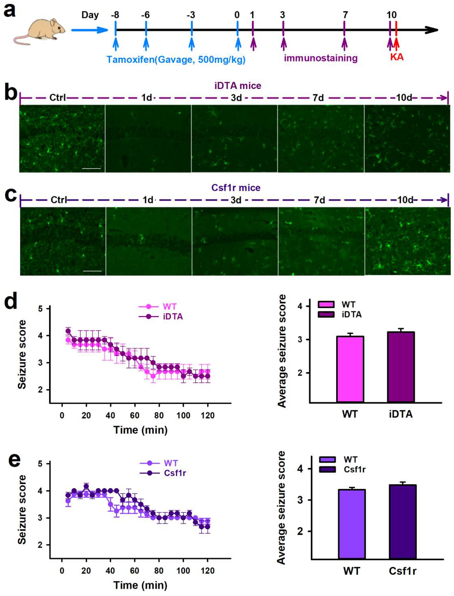
Microglia are the resident immune cells of the center nervous system and participate in various neurological diseases. Here we determined the function of microglia in epileptogenesis using microglial ablation approaches. Three different microglia-specific genetic tools were used, CX3CR1CreER/+:R26iDTA/+, CX3CR1CreER/+:R26iDTR/+, and CX3CR1CreER/+:Csf1rFlox/Flox mice. We found that microglial depletion led to worse kainic acid (KA)-induced status epilepticus, higher mortality rate, and increased neuronal degeneration in the hippocampus. In KA-induced chronic spontaneous recurrent seizures, microglial depletion increased seizure frequency, interictal spiking, and seizure duration. Therefore, microglial depletion aggravates the severity of KA-induced acute and chronic seizures. Interestingly, microglial repopulation reversed the effects of depletion upon KA-induced status epilepticus. Our results demonstrate a beneficial role of microglia in suppressing both acute and chronic seizures, suggesting that microglia are a potential therapeutic target for the management of epilepsy.

Keywords: Epilepsy; Kainic acid; Microglia; Microglia depletion; Microglia repopulation; Neuronal degeneration; Spontaneous recurrent seizures; Status epilepticus.


Conflict of interest statement

Declaration of Competing Interest The authors declare that they have no known competing financial interests or personal relationships that could have appeared to influence the work reported in this paper.

Figures

Figure 1.
Figure 1.. Microglia depletion and re-population in the mouse brain.
a, Tamoxifen treatment scheme. b,e, Representative images showing microglia depletion and re-population in the brain of iDTA mice (b) and Csf1r mice (e). Using Analyze Particles in ImageJ to perform automatic cell counting, each blue dot represents a microglial cell. c,f, Immunostaining images of Iba1+ microglial cells in the hippocampus and cortex of iDTA mice and Csf1r mice during the time course of depletion and re-population. Scale bar=50 μm. d,g, Quantification of Iba+ microglial cells in the brain of iDTA mice and Csf1r mice treated with tamoxifen. Data presented as means ± SEM, n=3–4, ***P<0.001 vs 0d.
Figure 2.
Figure 2.. Re-populated cells are P2Y12+ microglia.
a, The scheme of tamoxifen treatment. b,c Representative images show that CD11b (green) and P2Y12 (red) are co-expressed in re-populated cells in the brain of iDTA mice (b) and Csf1r mice (c). Scale bar=50 μm.
Figure 3.
Figure 3.. Microglia depletion increases acute seizure in response to KA treatment.
a, Drug treatment and behavior test scheme in iDTA and Csf1r mice. b–e, Quantification of seizure scores and mortality in iDTA mice (b–c) and Csf1r mice (d–e) after ICV KA. f, Drugs treatment and immunostaining scheme in iDTR mice. g, Representative images and quantification of Iba-1+ cells showing microglia depletion and re-population in iDTR mice at 1d, 2d, 3d, 5d, and 7d after tamoxifen and DT treatment. Scale bar=100 μm. h, Drugs treatment and behavior test scheme in iDTR mice. i–j, Quantification of seizure scores and mortality in iDTR mice after ICV KA. Data presented means ± SEM, n=6–8. ***P<0.001 or **P<0.01 vs control group.
Figure 4.
Figure 4.. Microglia depletion increases neuronal injury in response to KA treatment.
a, Drug treatment and immunostaining scheme in iDTA and Csf1r mice. b–e, Representative images and quantification of FJC+ neurons show that CX3CR1+ cell depletion worsens neuronal damage in the hippocampus of iDTA mice (b, c) and Csf1r mice (d, e) 1d and 3d after ICV KA treatment. f, Drug treatment and immunostaining scheme in iDTR mice. g–h, Representative images and quantification of FJC+ neurons show that specific microglia depletion worsens neuronal damage in the hippocampus of iDTR mice 1d and 3d after KA treatment. Scale bar=100 μm. Data presented as means ± SEM, n= 3, **P<0.01 vs control group.
Figure 5.
Figure 5.. Microglia depletion aggravates spontaneous seizure induced by KA.
a, Drug treatment and EEG recording scheme in iDTA and Csf1r mice. b–e, Representative traces and quantification of EEG recordings show that CX3CR1+ cell ablation aggravates spontaneous seizures in iDTA mice (b, c) and Csf1r mice (d, e) starting 28 days after KA treatment. f, Drug treatment and EEG recording scheme in iDTR mice. g–h, Representative traces and quantification of EEG recordings show that specific microglia depletion increases spontaneous seizures in iDTR mice starting 28 days after KA treatment. Data presented as means ± SEM, n=3–4. **P<0.01 vs control group.
Figure 6.
Figure 6.. Re-populated microglia did not alter seizures induced by KA.
a, Drug treatment, immunostaining and behavior test scheme in iDTA and Csf1r mice. b–c, Representative images showing the time course of microglia repopulation in the hippocampus following rapid depletion in iDTA mice (b) and Csf1r mice (c). Scale bar =50 μm, n=3. d–e, Quantification of seizure scores show that iDTA mice and Csf1r mice with repopulated microglia have similar seizure scores compared with their respective controls. Data presented as means ± SEM, n=6–8.

Comment in

References

    1. Abraham J, Fox PD, Condello C, Bartolini A, Koh S, 2012. Minocycline attenuates microglia activation and blocks the long-term epileptogenic effects of early-life seizures. Neurobiology of disease 46, 425–430. - PMC - PubMed
    1. Avignone E, Ulmann L, Levavasseur F, Rassendren F, Audinat E, 2008. Status epilepticus induces a particular microglial activation state characterized by enhanced purinergic signaling. J Neurosci 28, 9133–9144. - PMC - PubMed
    1. Baalman K, Marin MA, Ho TS, Godoy M, Cherian L, Robertson C, Rasband MN, 2015. Axon initial segment-associated microglia. J Neurosci 35, 2283–2292. - PMC - PubMed
    1. Bhandare AM, Kapoor K, Powell KL, Braine E, Casillas-Espinosa P, O’Brien TJ, Farnham MMJ, Pilowsky PM, 2017. Inhibition of microglial activation with minocycline at the intrathecal level attenuates sympathoexcitatory and proarrhythmogenic changes in rats with chronic temporal lobe epilepsy. Neuroscience 350, 23–38. - PubMed
    1. Brown GC, Neher JJ, 2010. Inflammatory neurodegeneration and mechanisms of microglial killing of neurons. Molecular neurobiology 41, 242–247. - PubMed

Publication types