. 2020 Jul 21;119(2):243-257.
doi: 10.1016/j.bpj.2020.05.036. Epub 2020 Jun 12.

Intercellular Adhesion Stiffness Moderates Cell Decoupling as a Function of Substrate Stiffness

Affiliations

Intercellular Adhesion Stiffness Moderates Cell Decoupling as a Function of Substrate Stiffness

Diego A Vargas et al. Biophys J. .

Abstract

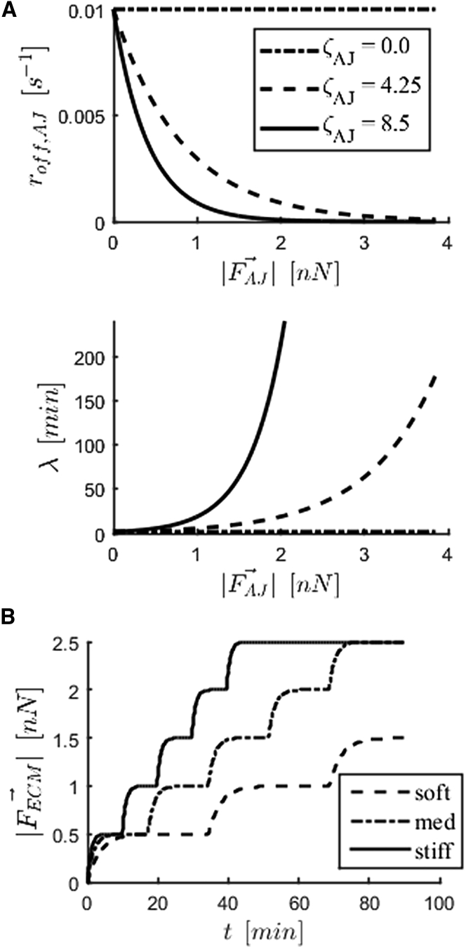
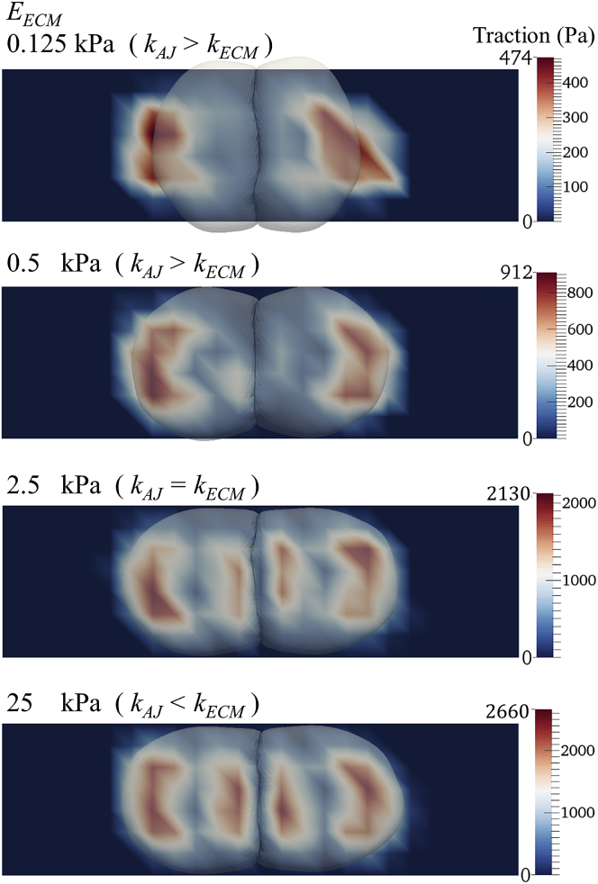
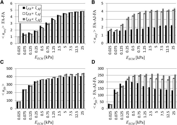
The interplay between cell-cell and cell-substrate interactions is complex yet necessary for the formation and healthy functioning of tissues. The same mechanosensing mechanisms used by the cell to sense its extracellular matrix also play a role in intercellular interactions. We used the discrete element method to develop a computational model of a deformable cell that includes subcellular components responsible for mechanosensing. We modeled a three-dimensional cell pair on a patterned (two-dimensional) substrate, a simple laboratory setup to study intercellular interactions. We explicitly modeled focal adhesions and adherens junctions. These mechanosensing adhesions matured, becoming stabilized by force. We also modeled contractile stress fibers that bind the discrete adhesions. The mechanosensing fibers strengthened upon stalling. Traction exerted on the substrate was used to generate traction maps (along the cell-substrate interface). These simulated maps are compared to experimental maps obtained via traction force microscopy. The model recreates the dependence on substrate stiffness of the tractions' spatial distribution, contractile moment of the cell pair, intercellular force, and number of focal adhesions. It also recreates the phenomenon of cell decoupling, in which cells exert forces separately when substrate stiffness increases. More importantly, the model provides viable molecular explanations for decoupling: mechanosensing mechanisms are responsible for competition between different fiber-adhesion configurations present in the cell pair. The point at which an increasing substrate stiffness becomes as high as that of the cell-cell interface is the tipping point at which configurations that favor cell-substrate adhesion dominate over those favoring cell-cell adhesion. This competition is responsible for decoupling.


Figures

Figure 1
Figure 1
Schematic of a cross section of cell pair displaying the different parts represented in the model (top). Forces involved in evolution of the mechanical system (bottom). The following relevant forces are indicated: cortical elastic spring (FLinear), cortical dissipation (FDashpot), cortical bending (Fbend), local triangle and global cell area conservation (FA), cell volume conservation (Fvolume), membrane contact (FMD), FA (FFA), AJ (FAJ), and stress fiber (FSF). To see this figure in color, go online.
Figure 2
Figure 2
Implementation of mechanosensing mechanisms in the cell. (A) AJ disassembly rate (roff,AJ) decreases with force carried by adhesion (|FAJ|)(top). Corresponding expected adhesion lifetimes (λ), where roff,AJ = 1 − e−1/2 (bottom). Parameter ζAJ dictates sensitivity to force. (B) Step increase in force carried by ECM (|FECM|) bound to an FA and applied by a stress fiber. Steps correspond to stalling in fiber contraction. The stiffer the substrate, the faster a fiber strengthens, taking more steps during its lifetime. Graphs shown are a result of a simulation of an isolated two-spring system (ideal case).
Figure 3
Figure 3
Traction maps for simulation of cell pairs on substrates of varying stiffness while maintaining constant AJ stiffness. Decoupling with increasing substrate stiffness (EECM) is evidenced by the spatial redistribution of cellular tractions in the cell-substrate interface from underneath the outer region of the cell pair to underneath the cell-cell interface. These sample cases correspond to simulations with maturing FAs, but not AJs (ζFA > ζAJ). An animation of the entire simulation for each of these cases can be found as Videos S1, S2, S3, and S4. To see this figure in color, go online.
Figure 4
Figure 4
For a Figure360 author presentation of this figure, see https://doi.org/10.1016/j.bpj.2020.05.036. The mechanosensing capability of adherens junctions relative to focal adhesions was varied by varying the parameter ζAJ while keeping ζFA the same. Average values for simulated cell pairs of (A) intercellular force (Fj,cell), (B) contractile moment of the cell pair (Mxx), (C) cell coupling (ψ), and (D) number of FAs bound to a stress fiber. n = 5. Error bars correspond to standard error of the mean. (E) Corresponding experimental results. Adapted from Polio et al. (6).
Figure 5
Figure 5
(A) Average value of factor describing stress fiber strengthening for fibers in cell pair connecting two FAs (<nstr,f> FA-FA). (B) Average value of factor describing stress fiber strengthening for fibers in cell pair connected to another via an AJ (<nstr,f> FA-AJ-FA). (C) The number of fibers in cell pair in all configurations (<nfib>). (D) The number of fibers in cell pair connected to another via an AJ(<nfib> FA-AJ-FA). n = 5. Error bars correspond to standard error of the mean.

Comment in

  • Stiffness Decouples Cellular Mechanosensation.
    Subramanianbalachandar V, Steward R Jr. Subramanianbalachandar V, et al. Biophys J. 2020 Jul 21;119(2):233. doi: 10.1016/j.bpj.2020.06.008. Epub 2020 Jun 12. Biophys J. 2020. PMID: 32610091 Free PMC article. No abstract available.

References

    1. Mui K.L., Chen C.S., Assoian R.K. The mechanical regulation of integrin-cadherin crosstalk organizes cells, signaling and forces. J. Cell Sci. 2016;129:1093–1100. - PMC - PubMed
    1. Matsushita T., Oyamada M., Takamatsu T. Remodeling of cell-cell and cell-extracellular matrix interactions at the border zone of rat myocardial infarcts. Circ. Res. 1999;85:1046–1055. - PubMed
    1. Araujo B.B., Dolhnikoff M., Mauad T. Extracellular matrix components and regulators in the airway smooth muscle in asthma. Eur. Respir. J. 2008;32:61–69. - PubMed
    1. An S.S., Mitzner W., Liggett S.B. An inflammation-independent contraction mechanophenotype of airway smooth muscle in asthma. J. Allergy Clin. Immunol. 2016;138:294–297.e4. - PMC - PubMed
    1. Tan C., Costello P., Dedhar S. Inhibition of integrin linked kinase (ILK) suppresses β-catenin-Lef/Tcf-dependent transcription and expression of the E-cadherin repressor, snail, in APC-/- human colon carcinoma cells. Oncogene. 2001;20:133–140. - PubMed

Publication types

LinkOut - more resources