. 2020 Jul 24;23(7):101288.
doi: 10.1016/j.isci.2020.101288. Epub 2020 Jun 20.

Bacterial Quorum Sensing Molecules Promote Allergic Airway Inflammation by Activating the Retinoic Acid Response

Affiliations

Bacterial Quorum Sensing Molecules Promote Allergic Airway Inflammation by Activating the Retinoic Acid Response

Renlan Wu et al. iScience. .

Abstract

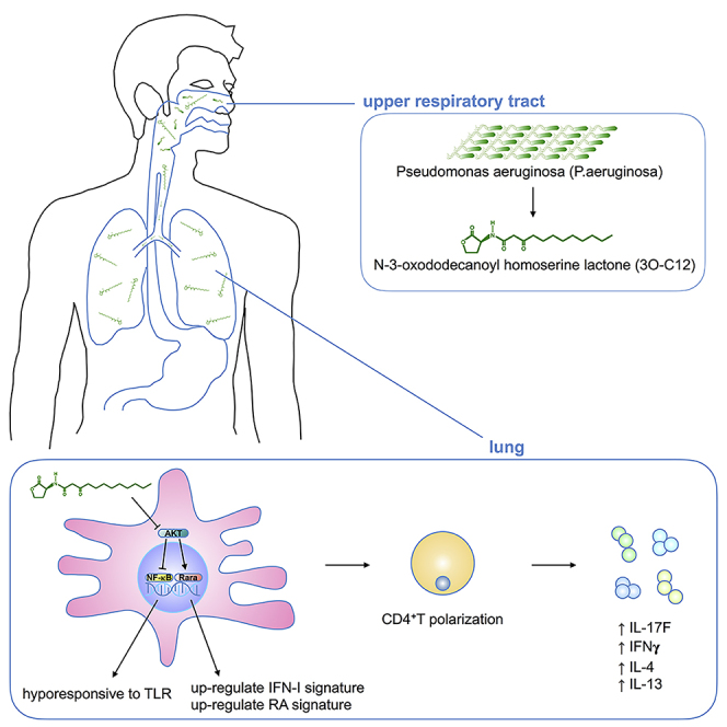
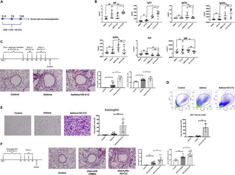
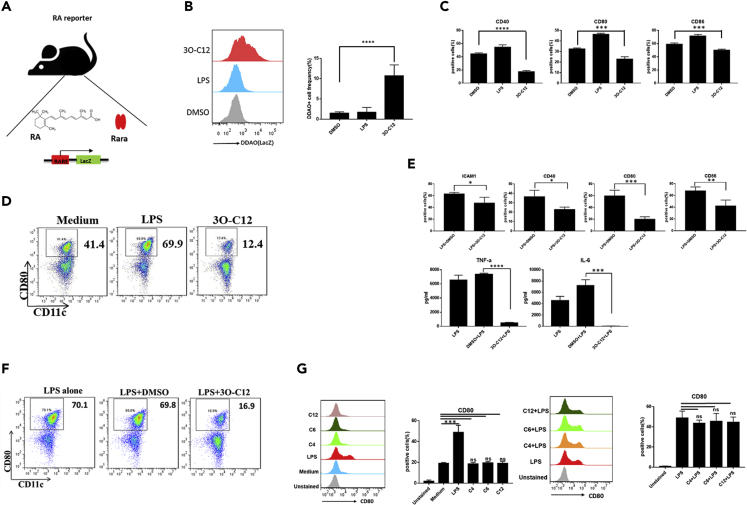
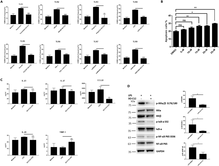
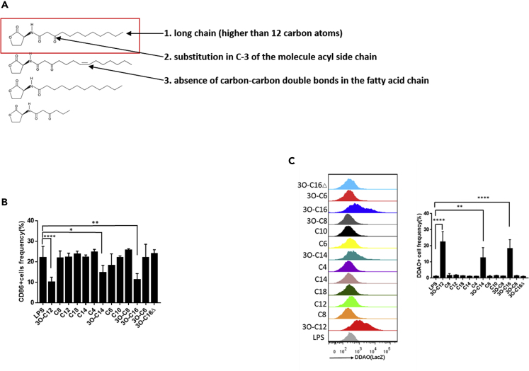
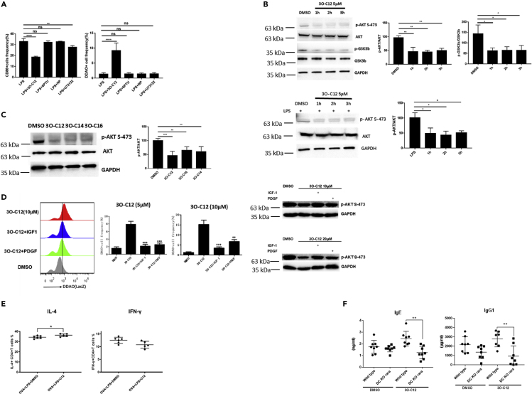
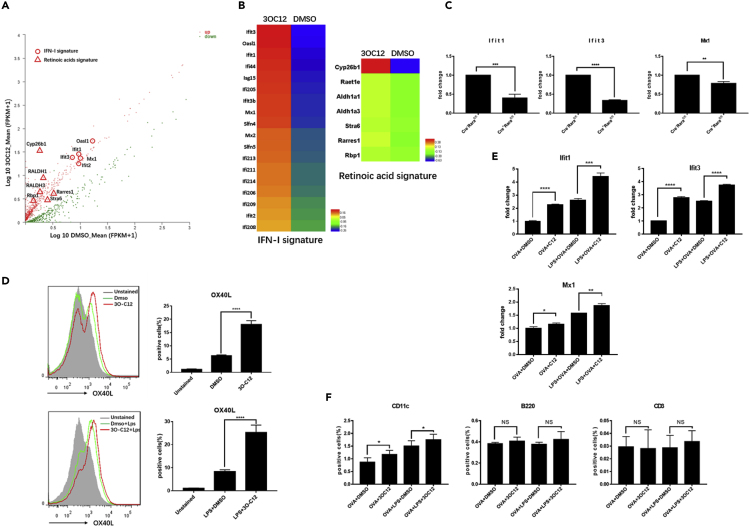
IgE and IgG1 production in the type 2 immune response is the characteristic feature of an allergic reaction. However, whether bacterial molecules modulate IgE and IgG1 production remains obscure. Here, we demonstrate that the bacterial quorum sensing molecules acyl homoserine lactones (AHLs) induce IgE and IgG1 production by activating the RARE (retinoic acid response element) response in dendritic cells (DCs) in vivo. DC-specific knockout of the retinoic acid transcriptional factor Rara diminished the AHL-stimulated type 2 immune response in vitro. AHLs altered DC phenotype, upregulated OX40L and IFN-I signature, and promoted T helper 2 cell differentiation in vitro. Finally, AHLs activated the RARE response by inhibiting AKT phosphorylation in vitro, as the AKT agonists IGF-1 and PDGF abolished the effect of AHLs on the RARE response. This study demonstrates a mechanism by which AHLs drive allergic airway inflammation through activating retinoic acid signaling in DCs.

Keywords: Biological Sciences; Immune Response; Immunology.


Conflict of interest statement

Declaration of Interests The authors declare no competing interests.

Figures

None
Graphical abstract
Figure 1
Figure 1
3O-C12 Stimulates OVA-Specific IgE and IgG1 Production and Enhances Allergic Lung Inflammation (A) Graphical representation of mouse immunization. (B) Wild-type C57BL/6 mice (6–8 weeks, male and female) were subcutaneously immunized with OVA (50 μg/mouse) + LPS (0.5 μg/mouse) with or without 3O-C12 (250 μg/mouse) every week for 3 weeks; incomplete Freund adjuvant was used as a vehicle. Mouse serum was collected; OVA-specific IgE, IgG1, IgG2a, IgG2c, IgM, IgA, and IgG2b were analyzed with ELISA. Results are representative of three independent experiments. (C–E) (C) BALB/c mice were divided into three groups: control (n = 4), asthma (n = 15), and asthma + 3O-C12 (n = 6). The mice were intraperitoneally (i.p.) injected on days 1 and 13 with OVA (50 μg) and aluminum hydroxide (2 mg). On days 20 and 22, the mice were intranasally (i.n.) challenged with OVA (40 μg) and i.p. administered with or without 3O-C12 (500 μg). On days 25, 27, and 29, the mice were i.n. challenged with OVA (40 μg) and i.n. administered with or without 3O-C12 (50 μg). One day after the last OVA challenge, mice were sacrificed and assessed for lung histology (scale bar, 50 μm) (C) and bronchoalveolar lavage fluid (BALF) total cell number (D) and eosinophil number (E). (F) BMDCs from C57BL/6 mice were stimulated by OVA (200 μg/mL) and LPS (100 ng/mL) overnight, with or without 3O-C12 (10 μM). BMDCs were transferred i.n. to recipients (C57BL/6 mice) (n = 3 in control group; n = 10 in OVA + LPS + DMSO group; n = 11 in OVA + LPS + 3O-C12 group) on days 1 and 12. Each recipient was challenged with 40 μg OVA i.n. on days 13, 14, and 15. One day after the last OVA challenge, mice were sacrificed and assessed for lung histology (scale bar, 50 μm). The statistical quantification (inflammatory cells and airway thickening) of Figures 1C and 1F was analyzed by ImageJ software. Data are presented as mean ± SD; p values were calculated using the two-tailed Student's t test. Data that did not exhibit a normal distribution were analyzed using the nonparametric Kruskal-Wallis test with Dunn's post hoc test. ∗p < 0.05, ∗∗p < 0.01, ∗∗∗p < 0.001, ∗∗∗∗p < 0.0001; n.s., not significant.
Figure 2
Figure 2
3O-C12 Activates the RARE Response (A) Graphical representation of the working model of RA reporter mice. (B) BMDCs (5–7 days) cultured from RA reporter mice were stimulated with 3O-C12 (10 μM) or LPS (100 ng/mL) for 1–2 h and incubated with DDAOG (10 μM). DDAO fluorescence was analyzed by flow cytometry. Data are representative of three independent experiments (n = 5–7). (C) BMDCs (5–7 days) cultured from RA reporter mice were stimulated with 3O-C12 (10 μM) or LPS (100 ng/mL) overnight. Expression levels of CD40, CD80, and CD86 were determined by flow cytometry; cells were gated on CD11c-positive cells. Data are representative of three independent experiments (n = 5–7). (D) DC Magnetic-activated cell sorting (MACS) isolated from the spleen of C57BL/6 mice were stimulated with LPS (100 ng) or 3O-C12 (10 μM) or were unstimulated (medium). CD80 expression was determined by flow cytometry. Data are representative of three independent experiments (n = 5–7). (E) BMDCs (5–7 days) cultured from RA reporter mice were stimulated with LPS (100 ng/mL) overnight, with or without 3O-C12 (10 μM). Expression levels of ICAM1, CD40, CD80, and CD86 were determined by flow cytometry. Cytokine levels of IL-6 and TNF-α were evaluated by ELISA. Data are representative of three independent experiments (n = 5–7). (F) DC MACS isolated from the spleen of C57BL/6 mice were stimulated with LPS (100 ng) with or without 3O-C12 (10 μM) overnight. CD80 expression was determined by flow cytometry. Data are representative of three independent experiments (n = 5–7). (G) BMDCs (5–7 days) cultured from RA reporter mice were stimulated with C4 (10 μM), C6 (10 μM), and C12 (10 μM) alone or plus LPS. CD80 expression was determined by flow cytometry. Data are representative of three independent experiments (n = 5–7). Data are presented as mean ± SD; p values were calculated using two-tailed Student's t test. ∗p < 0.05, ∗∗p < 0.01, ∗∗∗p < 0.001, ∗∗∗∗p < 0.0001; n.s., not significant.
Figure 3
Figure 3
3O-C12 Inhibits TLR-Induced DC Activation (A) BMDCs (5–7 days) cultured from C57BL/6 mice were stimulated with different TLR ligands with or without 3O-C12 (10 μM) overnight. CD86-positive cells were estimated by flow cytometry. Data are representative of three independent experiments (n = 5–7). (B) BMDCs (5–7 days) cultured from C57BL/6 mice were incubated with different doses of 3O-C12 for 12 h; annexin V-positive cells were determined by flow cytometry. Data are representative of three independent experiments (n = 5–7). (C) BMDCs (5–7 days) cultured from C57BL/6 mice were unstimulated (medium) or stimulated with LPS or LPS + 3O-C12 (5 μM) overnight. Cytokines were determined by MILLIPLEX Multiplex Assays. Data are representative of three independent experiments (n = 5–7). (D) BMDCs (5–7 days) cultured from C57BL/6 mice were incubated with LPS plus 3O-C12 (10 μM) for 3 h. Cells were collected and lysed and activation of the NF-κB pathway was determined by western blot (n = 4). Data are presented as mean ± SD; p values were calculated using the two-tailed Student's t test. ∗p < 0.05, ∗∗p < 0.01, ∗∗∗∗p < 0.0001; n.s., not significant.
Figure 4
Figure 4
The Chemical Structure of AHLs Determines the RARE Response (A) The RARE response depends on the chemical structure of AHLs with three characteristics. (B) BMDCs (5–7 days) cultured from C57BL/6 mice were stimulated with LPS (100 ng/mL) with or without AHLs (10 μM) overnight. CD86-positive cells were estimated by flow cytometry. Data are representative of three independent experiments (n = 5–7). (C) BMDCs (5–7 days) cultured from RA reporter mice were stimulated with AHLs (10 μM) or LPS (100 ng/mL) for 1–2 h and incubated with DDAOG (10 μM) for 1–2 h. DDAO fluorescence was analyzed by flow cytometry. Data are representative of three independent experiments (n = 5–7). Data are presented as mean ± SD; p values were calculated using the two-tailed Student's t-test. ∗p < 0.05, ∗∗p < 0.01, ∗∗∗∗p < 0.0001; n.s., not significant.
Figure 5
Figure 5
3O-C12 Activates the RARE Response by Disrupting AKT Signaling (A) BMDCs (5–7 days) cultured from RA reporter mice were stimulated with LPS alone (100 ng/mL) or LPS plus 3O-C12 (33 μM), 6PTU (100 uM), NP (100 uM), or U73122 (10 μM) overnight and incubated with DDAOG (10 μM) for 1–2 h. DDAO fluorescence and CD86 expression were analyzed by flow cytometry. Data are representative of three independent experiments. (B and C) (B) BMDCs (5–7 days) cultured from C57BL/6 mice were incubated with 3O-C12 (10 μM) for 1–3 h or (C) LPS plus 3O-C12. Cells were collected and lysed, and AKT phosphorylation was determined by western blot. Results are representative of three independent experiments. (D) BMDCs (5–7 days) cultured from RA reporter mice were stimulated with 3O-C12 (5 μM and 10 μM) or 3O-C12 plus IGF-1 (200 ng/mL) and PDGF (40 ng/mL) for 1–2 h and incubated with DDAOG (10 μM) for 1–2 h. DDAO fluorescence was analyzed by flow cytometry. AKT phosphorylation was determined by western blot. Data are representative of three independent experiments. (E) BMDCs (5–7 days) cultured from C57BL/6 mice were loaded with OVA (100 μg/mL), incubated with LPS, or LPS+3O-C12 (10 μM) for 1–3 h; naive OT-II cells were added and co-cultured for 5 days. IL4- and IFN-γ-positive cells were determined by flow cytometry. Data are representative of three independent experiments. (F) 6- to 8-week-old male and female CD11ccre+Rarafl/fl mice (DC KO Rara) and littermate control CD11ccre-Rarafl/fl mice (wild-type) were subcutaneously immunized with OVA (50 μg/mice) + LPS (0.5 μg/mice) with or without 3O-C12 (250 μg/mouse) every week for 3 weeks. Mouse serum was collected, and OVA-specific IgE and IgG1 were analyzed by ELISA. Results are representative of three independent experiments. Data are presented as mean ± SD; p values were calculated using the two-tailed Student's t test. ∗p < 0.05, ∗∗p < 0.01, ∗∗∗p < 0.001, ∗∗∗∗p < 0.0001; n.s., not significant.
Figure 6
Figure 6
3O-C12 Stimulates the IFN-I Signature and OX40L (A and B) RNA sequencing analysis of the gene expression of 3O-C12-activated DCs. (A) Scatterplot of gene expression of FPKM values. (B) Heatmap of FPKM values, IFN-I signature, and RA signature. (C) The 3O-C12-stimulated IFN-I signature depends on Rara. Gene expression levels were examined by qPCR. Results are representative of two independent experiments. (D) Fluorescence-activated cell sorting analysis of OX40L in 3O-C12-activated DCs (upper panel) and 3O-C12+LPS-activated DCs (lower panel). Results are representative of four independent experiments. (E) 3O-C12 stimulates expression of the IFN-I signature in DCs in vivo. Results are representative of three independent experiments. (F) 3O-C12 upregulates OX40L in DCs in vivo. Results are representative of two independent experiments (n = 5–7). p values were calculated using the two-tailed Student's t test. ∗p < 0.05, ∗∗p < 0.01, ∗∗∗p < 0.001, ∗∗∗∗p < 0.0001; n.s., not significant.

Similar articles

Cited by

References

    1. Bachus H., Kaur K., Papillion A.M., Marquez-Lago T.T., Yu Z., Ballesteros-Tato A., Matalon S., Leon B. Impaired tumor-necrosis-factor-alpha-driven dendritic cell activation limits lipopolysaccharide-induced protection from allergic inflammation in infants. Immunity. 2019;50:225–240.e4. - PMC - PubMed
    1. Bhattacharya N., Yuan R., Prestwood T.R., Penny H.L., DiMaio M.A., Reticker-Flynn N.E., Krois C.R., Kenkel J.A., Pham T.D., Carmi Y. Normalizing microbiota-induced retinoic acid deficiency stimulates protective CD8(+) T cell-mediated immunity in colorectal cancer. Immunity. 2016;45:641–655. - PMC - PubMed
    1. Bisgaard H., Li N., Bonnelykke K., Chawes B.L., Skov T., Paludan-Muller G., Stokholm J., Smith B., Krogfelt K.A. Reduced diversity of the intestinal microbiota during infancy is associated with increased risk of allergic disease at school age. J. Allergy Clin. Immunol. 2011;128:646–652.e1-5. - PubMed
    1. Cait A., Hughes M.R., Antignano F., Cait J., Dimitriu P.A., Maas K.R., Reynolds L.A., Hacker L., Mohr J., Finlay B.B. Microbiome-driven allergic lung inflammation is ameliorated by short-chain fatty acids. Mucosal Immunol. 2018;11:785–795. - PubMed
    1. Chen C., Perry T.L., Chitko-McKown C.G., Smith A.D., Cheung L., Beshah E., Urban J.F., Jr., Dawson H.D. The regulatory actions of retinoic acid on M2 polarization of porcine macrophages. Dev. Comp. Immunol. 2019;98:20–33. - PubMed

LinkOut - more resources