

Spectrum of chest computed tomographic (CT) findings in coronavirus disease-19 (COVID-19) patients in India
- PMID: 32623113
- PMCID: PMC7313528
- DOI: 10.1016/j.ejrad.2020.109147
Spectrum of chest computed tomographic (CT) findings in coronavirus disease-19 (COVID-19) patients in India
Abstract
Purpose: To report the spectrum of chest computed tomographic (CT) imaging findings in coronavirus disease-19 (COVID-19) infected Indian patients.
Methods: This was a prospective descriptive study comprising 147 consecutive reverse transcriptase polymerase chain reaction (RT-PCR) positive patients who underwent CT chest. Prevalence, distribution, extent and type of abnormal lung findings were recorded.
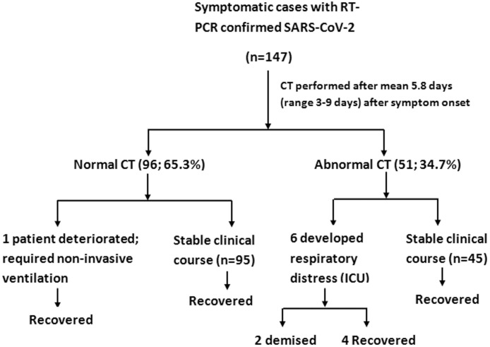
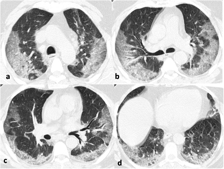
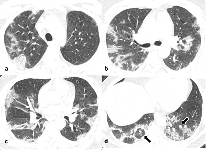
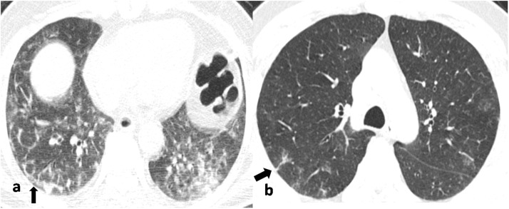
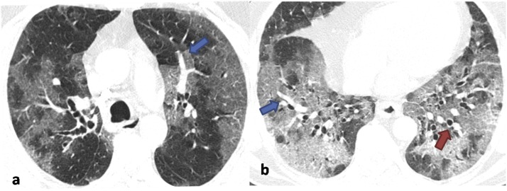
Results: Among the total study cohort of 147 patients, 104 (70.7 %) were males and 43 (29.3 %) were females with mean age of 40.9 ± 17.2 years (range 24-71 years). We observed lung parenchymal abnormalities in 51 (34.7 %) cases whereas 96 (65.3 %) RT-PCR positive cases had a normal chest CT. Only 12.2 % of the patients were dyspneic, 6.1 % had desaturation, 7.4 % had increased respiratory rate and 10.9 % had comorbidities. Among the patients with abnormal CT findings bilateral 39/51 (76.5 %), multilobar (88.2 %) lung involvement with a predominant peripheral and posterior distribution was commonly observed. With regards to the type of opacity, ground glass opacity (GGO) was the dominant abnormality found in all 51 (100 %) cases. Pure GGO was observed in 15 (29.4 %), GGO with crazy paving pattern was seen in 15 (29.4 %) and GGO mixed with consolidation was noted in 21(41.2 %). Peri-lesional or intralesional segmental or subsegmental pulmonary vessel enlargement was observed in 36 (70.6 %) cases.
Conclusion: In this study population predominantly with mild symptoms and few comorbidities, two-thirds of RT-PCR positive patients had a normal chest CT; whereas the remaining patients showed typical findings of predominant GGOs with a bilateral distribution and peripheral predominance.
Keywords: COVID-19; CT; SARS-CoV-2.
Copyright © 2020 Elsevier B.V. All rights reserved.
Conflict of interest statement
Declaration of Competing Interest None.
Figures
References
-
- World Health Organization . 2020. Coronavirus Disease 2019 (COVID-19): Situation Report. 137.
-
- Kong W.H., Li Y., Peng M.W., Kong D.G., Yang X.B., Wang L., Liu M.Q. SARS-CoV-2 detection in patients with influenza-like illness. Nat. Microbiol. 2020;(April):1–4. - PubMed
-
- Yang Y., Yang M., Shen C. 2020. Evaluating the Accuracy of Different Respiratory Specimens in the Laboratory Diagnosis and Monitoring the Viral Shedding of 2019-nCoV Infections. - DOI
Publication types
MeSH terms
LinkOut - more resources
Full Text Sources
Medical
Miscellaneous
