. 2020:27:102323.
doi: 10.1016/j.nicl.2020.102323. Epub 2020 Jun 24.

Characterization of serial hyperpolarized 13C metabolic imaging in patients with glioma

Affiliations

Characterization of serial hyperpolarized 13C metabolic imaging in patients with glioma

Adam W Autry et al. Neuroimage Clin. 2020.

Abstract

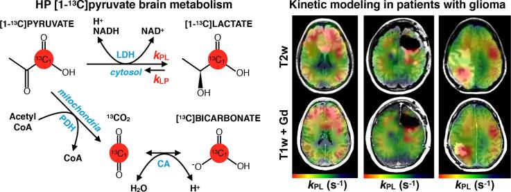
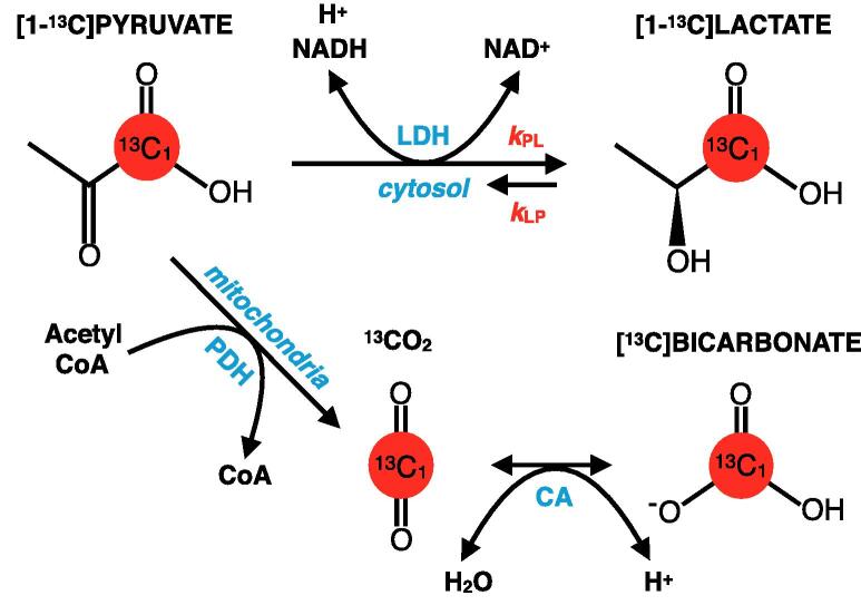
Background: Hyperpolarized carbon-13 (HP-13C) MRI is a non-invasive imaging technique for probing brain metabolism, which may improve clinical cancer surveillance. This work aimed to characterize the consistency of serial HP-13C imaging in patients undergoing treatment for brain tumors and determine whether there is evidence of aberrant metabolism in the tumor lesion compared to normal-appearing tissue.

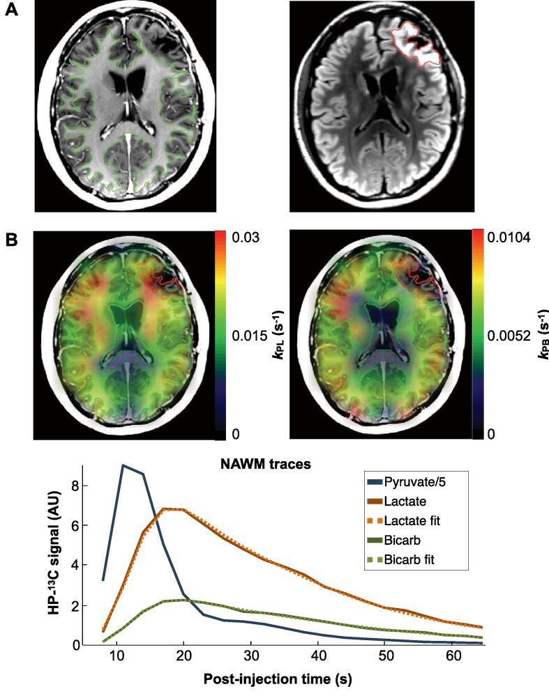
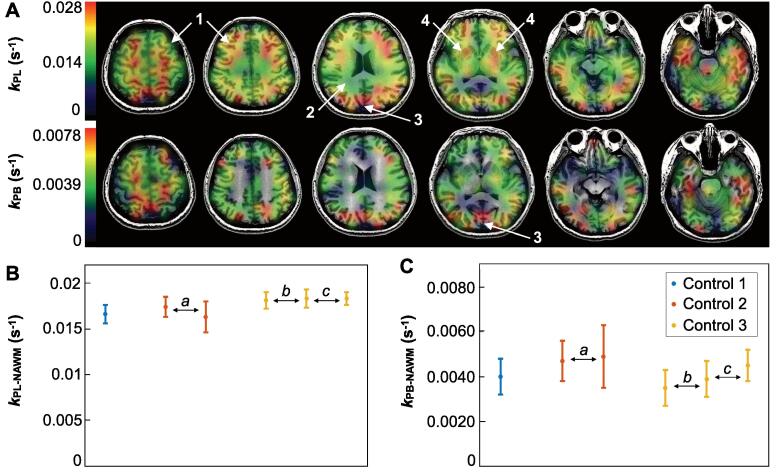
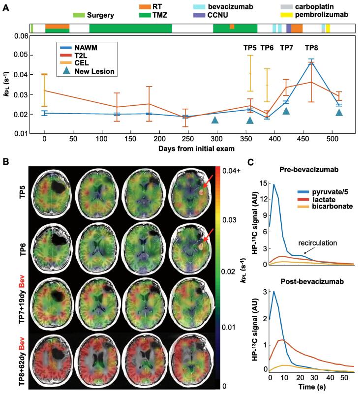
Methods: Serial dynamic HP [1-13C]pyruvate MRI was performed on 3 healthy volunteers (6 total examinations) and 5 patients (21 total examinations) with diffuse infiltrating glioma during their course of treatment, using a frequency-selective echo-planar imaging (EPI) sequence. HP-13C imaging at routine clinical timepoints overlapped treatment, including radiotherapy (RT), temozolomide (TMZ) chemotherapy, and anti-angiogenic/investigational agents. Apparent rate constants for [1-13C]pyruvate conversion to [1-13C]lactate (kPL) and [13C]bicarbonate (kPB) were simultaneously quantified based on an inputless kinetic model within normal-appearing white matter (NAWM) and anatomic lesions defined from 1H MRI. The inter/intra-subject consistency of kPL-NAWM and kPB-NAWM was measured in terms of the coefficient of variation (CV).

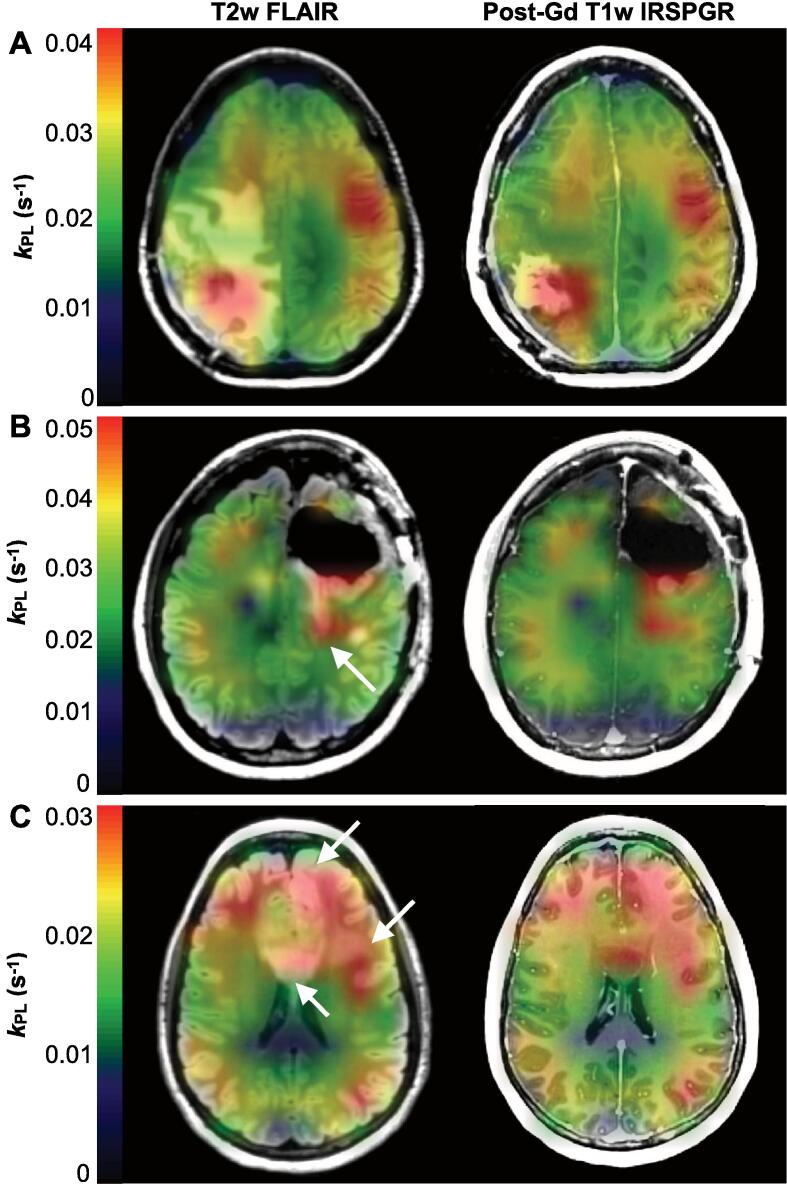
Results: When excluding scans following anti-angiogenic therapy, patient values of kPL-NAWM and kPB-NAWM were 0.020 s-1 ± 23.8% and 0.0058 s-1 ± 27.7% (mean ± CV) across 17 HP-13C MRIs, with intra-patient serial kPL-NAWM/kPB-NAWM CVs ranging 6.8-16.6%/10.6-40.7%. In 4/5 patients, these values (0.018 s-1 ± 13.4% and 0.0058 s-1 ± 24.4%; n = 13) were more similar to those from healthy volunteers (0.018 s-1 ± 5.0% and 0.0043 s-1 ± 12.6%; n = 6) (mean ± CV). The anti-angiogenic agent bevacizumab was associated with global elevations in apparent rate constants, with maximum kPL-NAWM in 2 patients reaching 0.047 ± 0.001 and 0.047 ± 0.003 s-1 (±model error). In 3 patients with progressive disease, anatomic lesions showed elevated kPL relative to kPL-NAWM of 0.024 ± 0.001 s-1 (±model error) in the absence of gadolinium enhancement, and 0.032 ± 0.008, 0.040 ± 0.003 and 0.041 ± 0.009 s-1 with gadolinium enhancement. The lesion kPB in patients was reduced to unquantifiable values compared to kPB-NAWM.

Conclusion: Serial measures of HP [1-13C]pyruvate metabolism displayed consistency in the NAWM of healthy volunteers and patients. Both kPL and kPB were globally elevated following bevacizumab treatment, while progressive disease demonstrated elevated kPL in gadolinium-enhancing and non-enhancing lesions. Larger prospective studies with homogeneous patient populations are planned to evaluate metabolic changes following treatment.

Keywords: Bevacizumab; Carbon-13; Glioma; Hyperpolarized; Kinetics; Metabolism.


Figures

None
Graphical abstract
Fig. 1
Fig. 1
HP [1-13C]pyruvate brain metabolism. Diagram of HP [1-13C]pyruvate metabolism in the brain, which is characterized by two primary pathways: enzymatic conversion of [1-13C]pyruvate to [1-13C]lactate via cytosolic lactate dehydrogenase (LDH); and successive conversion of [1-13C]pyruvate to 13CO2 and [13C]bicarbonate via mitochondrial pyruvate dehydrogenase (PDH) and carbonic anhydrase (CA), respectively. The second-order kinetics of pyruvate-to-bicarbonate conversion are approximated by the rate-limiting step of PDH, given the rapid CO2-bicarbonate exchange catalyzed by CA. HP [1-13C]pyruvate is also reversibly converted to [1-13C]alanine via alanine transaminase (ALT), but prior studies have shown that conversion to HP [1-13C]alanine occurs outside of the brain (4).
Fig. 2
Fig. 2
Example HP-13C kinetic maps. Regions of interest from a patient with GBM: NAWM (green) and T2L (red) overlaid on T1-weighted IRSPGR and T2-weighted FLAIR images, respectively (A). Maps of kPL and kPB based on kinetic modeling of dynamic HP-13C EPI data overlaid on the same T1-weighted images (B, top). Corresponding dynamic traces of HP [1-13C]pyruvate, [1-13C]lactate, and [13C]bicarbonate signal within NAWM are shown alongside kinetic model fits (B, bottom). (For interpretation of the references to colour in this figure legend, the reader is referred to the web version of this article.)
Fig. 3
Fig. 3
Volunteer HP-13C kinetic data. Maps of kPL and kPB from the third scan of healthy volunteer HV3 overlaid on T1-weighted images, which illustrate the spatial variation of apparent HP [1-13C]pyruvate metabolism: 1, cortex/grey matter; 2, white matter; 3, cuneus; and 4, putamen/deep grey matter (A). Healthy volunteer values of kPL-NAWM (B) and kPB-NAWM (C) are shown together with nonlinear least squares fitting error for 3 subjects over scan intervals of 30 min (a), 107 days (b) and 67 days (c).
Fig. 4
Fig. 4
Effects of bevacizumab. Serial kPL data within NAWM and presumed tumor regions are shown for patient P2 over 9 scans spanning 512 days, along with clinical treatment information (A). Values of kPL-NAWM (blue) remained consistent until the administration of bevacizumab, whereupon a global increase in kPL occurred, as seen at timepoint 8 (TP8) (A). Both kPL-T2L (red) and kPL-CEL (orange) are seen to be elevated relative to kPL-NAWM, particularly at the time of progression. Corresponding kPL maps for timepoints TP5-TP8 overlaid on T1-weighted images illustrate the emergence of a new gadolinium-enhancing lesion with elevated kPL (red arrows), which disappeared following treatment with bevacizumab, and subsequent global elevation of kPL (B). Kinetic traces from pre- and post-bevacizumab scans demonstrate lower overall HP signal with bevacizumab, but proportionally greater [1-13C]lactate and [13C]bicarbonate signal relative to that of [1-13C]pyruvate (C). (For interpretation of the references to colour in this figure legend, the reader is referred to the web version of this article.)
Fig. 5
Fig. 5
Profiles of progression. Imaging at the time of radiologically-defined progression shown using kPL maps overlaid on T2-weighted FLAIR and post-gadolinium T1-weighted images. Patient P5 demonstrated elevated kPL in the lesion that was spatially heterogeneous, with higher values in and around the CEL compared to the surrounding T2L (A); whereas patient P2 showed uniformly elevated kPL that extended distally from the CEL into the T2L, as indicated by the white arrow (B). Diffusely elevated kPL in patient P4 corresponded with a large non-enhancing T2L centered in the corpus callosum and extending to the left frontal white matter and cortex (white arrows) (C). In each case, the lesion kPL highlighted radiological progression.

References

    1. Ardenkjaer-Larsen J.H., Fridlund B., Gram A. Increase in signal-to-noise ratio of >10,000 times in liquid-state NMR. PNAS. 2003;100:10158–10163. - PMC - PubMed
    1. Autry A.W., Gordon J.W., Carvajal L., Mareyam H., Chen H.Y., Park I., Mammoli D., Vareth M., Chang S.M., Wald L.L., Xu D., Vigneron D.B., Nelson S.J., Li Y. Comparison between 8- and 32-channel phased-array receive coils for in vivo hyperpolarized 13 C imaging of the human brain. Magn. Reson. Med. 2019;82:833–841. - PMC - PubMed
    1. Bankson J.A., Walker C.M., Ramirez M.S. Kinetic modeling and constrained reconstruction of hyperpolarized [1-13C]-pyruvate offers improved metabolic imaging of tumors. Cancer Res. 2015;75(22):4708–4717. - PMC - PubMed
    1. Bastin M.E., Carpenter T.K., Armitage P.A., Sinha S., Wardlaw J.M., Whittle I.R. Effects of dexamethasone on cerebral perfusion and water diffusion in patients with high-grade glioma. AJNR. 2006;27(2):402–408. - PMC - PubMed
    1. Bian W., Li Y., Crane J.C., Nelson S.J.N. A fully automated atlas based method for prescribing 3D PRESS MR spectroscopic imaging: towards robust and reproducible metabolite measurements in human brain. Magn. Reson. Med. 2018;79(2):636–642. - PMC - PubMed

Publication types