. 2021 Feb;78(4):1501-1522.
doi: 10.1007/s00018-020-03580-1. Epub 2020 Jul 4.

Protein structure analysis of the interactions between SARS-CoV-2 spike protein and the human ACE2 receptor: from conformational changes to novel neutralizing antibodies

Affiliations

Protein structure analysis of the interactions between SARS-CoV-2 spike protein and the human ACE2 receptor: from conformational changes to novel neutralizing antibodies

Ivan Mercurio et al. Cell Mol Life Sci. 2021 Feb.

Abstract

The recent severe acute respiratory syndrome, known as Coronavirus Disease 2019 (COVID-19) has spread so much rapidly and severely to induce World Health Organization (WHO) to declare a state of emergency over the new coronavirus SARS-CoV-2 pandemic. While several countries have chosen the almost complete lock-down for slowing down SARS-CoV-2 spread, the scientific community is called to respond to the devastating outbreak by identifying new tools for diagnosis and treatment of the dangerous COVID-19. With this aim, we performed an in silico comparative modeling analysis, which allows gaining new insights into the main conformational changes occurring in the SARS-CoV-2 spike protein, at the level of the receptor-binding domain (RBD), along interactions with human cells angiotensin-converting enzyme 2 (ACE2) receptor, that favor human cell invasion. Furthermore, our analysis provides (1) an ideal pipeline to identify already characterized antibodies that might target SARS-CoV-2 spike RBD, aiming to prevent interactions with the human ACE2, and (2) instructions for building new possible neutralizing antibodies, according to chemical/physical space restraints and complementary determining regions (CDR) mutagenesis of the identified existing antibodies. The proposed antibodies show in silico high affinity for SARS-CoV-2 spike RBD and can be used as reference antibodies also for building new high-affinity antibodies against present and future coronaviruses able to invade human cells through interactions of their spike proteins with the human ACE2. More in general, our analysis provides indications for the set-up of the right biological molecular context for investigating spike RBD-ACE2 interactions for the development of new vaccines, diagnostic kits, and other treatments based on the targeting of SARS-CoV-2 spike protein.

Keywords: ACE2 and ACE inhibitors; COVID-19; Comparative modeling; Coronavirus; Fold recognition tools; Neutralizing antibodies; Receptor binding domain; SARS-CoV-2; Spike; Spike post-fusion conformation; n-CoV19.


Figures
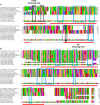
Fig. 1
Fig. 1
Extract of the MSA of SARS-CoV-2 spike protein monomer with the sequences of the crystallized structures of the spike whole protein or protein fragments observed in the post-fusion conformation from other coronaviruses, resulting from sequence cleavage. a Grey boxes indicate protein regions involved in cleavage events, whereas black boxes indicate the position of the proposed cleavage sites. a, b Cyan boxes indicate the cluster of residues conserved in the sampled sequences and maintained in the post-fusion conformation, considered as anchor points for preparing the 6b3o.pdb based SARS-CoV-2 spike sequence-structure alignment, used for building the 3D comparative model of SARS-CoV-2 spike protein in post-fusion conformation (amino acids S704-A771, see a; amino acids L922-S1147, see b; YP_009724390.1 residues numbering)
Fig. 2
Fig. 2
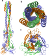
SARS-CoV-2 spike protein (S-II domain) 3D model in post-fusion conformation. Lateral view (a), top view (b) and bottom view (c) of the SARS-CoV-2 spike protein trimer 3D comparative model, reported in cartoon colored representation
Fig. 3
Fig. 3
SARS-CoV-2 spike protein regions involved in the pre/post-fusion conformational transitions. Topology panel: schematic of the SARS-CoV-2 spike protein organization according to the below reported 3D model protein regions. Colors and residues numbering reflect the localization in the SARS-CoV-2 spike protein sequence of the below reported protein domain 3D structures (YP_009724390.1 residues numbering). UH upstream helix; FP the region hosting the fusion peptide; HR1 heptad repeat 1; CH central helix; BH β-hairpin region; CD connector domain; HR2 heptad repeat 2, according to [45]. a, f Lateral views of the SARS-CoV-2 spike protein trimer in pre-/post-fusion conformation, respectively, are reported in colored cartoon representation. b, g Lateral views of the SARS-CoV-2 spike monomer in pre-/post-fusion conformation, respectively, are reported in colored cartoons. c, h Indicate the zoomed views of the Q926-K1028 protein region (green/red cartoon representation). d, i Indicate the zoomed views of the M1029-D1146 protein region (magenta/orange cartoon representation). e, j Indicate the zoomed views of the S704-I771 protein region (yellow cartoon representation). Yellow sticks in d, e, i, j indicate disulfide bridges. Residue labels indicate residues to be used as a reference for identifying quickly the cited protein region terminal portions or cysteine residues involved in disulfide bridges. Notably, yellow, magenta and red cartoon indicate the monomer regions involved in few conformational changes, whereas green and orange cartoon indicate regions involved in large conformational changes. Black cartoon portions in b indicate protein regions lost after cleavage events and/or not available in the crystallized structures. Cyan cartoon portion in g indicates the 1146–1197 protein region and was obtained by comparative modeling using as a protein template the only available SARS-CoV-1 spike protein with a solved structure for the corresponding protein region in the post-fusion conformation, as observed in 6b3o.pdb. Notably, the corresponding protein region was solved in none of the investigated crystallized spike proteins in pre-fusion conformations (Supplementary Table 1)
Fig. 4
Fig. 4
SARS-CoV-2 spike protein regions involved in the pre/post-fusion molecular packing. a, g Lateral views of the spike protein trimer in post/pre-fusion conformation, respectively. Colored regions indicate the main protein portions responsible for the different molecular packing of the spike protein in the post/pre-fusion conformations. b, h Lateral zoomed views of the spike protein trimer core (red–green–orange cartoon) in the post/pre-fusion conformations. c, e Top and bottom views of the spike protein trimer in post-fusion conformation reported in a. d, f Top and bottom views of the spike protein trimer colored regions in post-fusion conformation reported in b. i, k Top and bottom views of the spike protein trimer in pre-fusion conformation reported in g. j, l Top and bottom views of the spike protein trimer colored regions in post-fusion conformation reported in h. The reported colors indicate the same regions described in Fig. 3
Fig. 5
Fig. 5
Multiple sequence alignment of RBDs from 11 SARS-CoV and 3 MERS-CoV strains. The reported residues numbering refers to the indicated sequences sampled by blastp or to the indicated crystallized structure sequences
Fig. 6
Fig. 6
Side view (af) and top view (gl) of the human  SARS-CoV-2 spike protein interacting with 3 units of the human ACE2 N-terminal domain (ae; gk). SARS-CoV-2 spike protein trimer (6vsb.pdb) is reported in white cartoon representation with the 3 spike  RBDs reported in red (in the closed pre-fusion state) or green (in the open pre-fusion state) cartoon. The open pre-fusion state allows establishing pre-invasion interactions with the ACE2 N-terminal domain. SARS-CoV-2 spike protein trimer C-terminal domain, resulting from protein cleavage that triggers the post-fusion conformation, is reported in black cartoon representation in f (lateral view) and l (top view)
Fig. 7
Fig. 7
SARS-CoV-1 and SARS-CoV-2 RBD residues involved in direct interactions with ACE2. H. sapiens ACE2 is reported in white cartoon representation. SARS-CoV-1 RBD is reported in magenta cartoon representation, whereas SARS-CoV-2 RBD is reported in yellow cartoon representation. a, c Residues involved in polar interactions between SARS-CoV-1 RBD (magenta sticks) and ACE2 (white sticks). b, d Residues involved in polar interactions between SARS-CoV-2 RBD (yellow sticks) and ACE2 (black sticks). Polar interactions are represented by black dashed lines in the exploded views reported in c and d
Fig. 8
Fig. 8
SARS-CoV-1 spike and SARS-CoV-2 spike monomers in pre-fusion conformation interacting with SARS-CoV-1 spike RBD selective antibodies S230 (6nb7.pdb) and m396 (2dd8.pdb). a Superimposition of the tertiary structure of SARS-CoV-1 (6nb7.pdb) and SARS-CoV-2 (6vsb.pdb) spike protein monomers reported in pink cartoon representation. SARS-CoV-1 and SARS-CoV-2 RBDs are reported in grey cartoon representation. S230 FAB ab portion (6nb7.pdb) is reported in yellow (light chain) and pink (heavy chain) cartoon representation. m396 FAB ab portion (2dd8.pdb) is reported in orange (light chain) and blue (heavy chain) cartoon representation. b Zoomed view of the superimposition of SARS-CoV-1 Spike and SARS-CoV-2 Spike RBD domains interacting with S230 and m396 FAB antibodies (see a for colors). c, d Super zoomed and rotated views of the crystallized SARS-CoV-1 Spike RBD residues interacting with S230 ab. e, f Super zoomed and rotated views of SARS-CoV-2 Spike RBD predicted residues interacting with S230 ab. g, h Super zoomed and rotated views of the crystallized SARS-CoV-1 Spike RBD residues interacting with m396 ab. i, j Super zoomed and rotated views of SARS-CoV-2 Spike RBD predicted residues interacting with m396 ab. cj Residues at the RBD–ab interface in the 3.5–4 Å distance range are reported in sticks representation. White sticks indicate RBD residues; orange and blue sticks indicate m396 ab residues, yellow and pink sticks indicate S230 ab residues
Fig. 9
Fig. 9
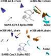
Molecular framework of the investigated proteins hosting SARS-CoV-spike RBDs, light and heavy chain of the m396 antibody and the human ACE2, simultaneously. The shown spike RBD, ACE2, and m396 protein portions are those in a reciprocal distance range of 4 Å. Upper panel: superimposition of the crystallized SARS-CoV-1 spike RBD (white cartoon representation) in complex with m396 antibody (2d88.pdb, orange, and blue cartoon) and ACE2 (2ajf.pdb, cyan cartoon). Bottom panel: superimposition of SARS-CoV-2 spike RBDs (from 6vw1.pdb, white cartoon representation), ACE2 from 6vw1.pdb (cyan cartoon) and m396 from 2d88.pdb (orange and blue cartoon)
Fig. 10
Fig. 10
m396 neutralizing antibody, native and engineered, in complex with SARS-CoV-2 spike RBD. a, b Exploded view and perspective view of native m396 neutralizing antibody (in orange blue cartoon) in complex with SARS-CoV-2 spike RBD (in white cartoon representation). Residues at the m396/RBD interface in a distance range within 4 Å are indicated by white sticks (RBD), orange sticks (m396 CDR-H residues), and blue sticks (m396 CDR-L residues). c, d Exploded view and perspective view of the engineered m396 predicted neutralizing antibody (in orange blue cartoon) in complex with SARS-CoV-2 spike RBD (in white cartoon representation). Residues at the engineered m396/RBD interface in a distance range within 4 Å are indicated by white sticks (RBD), orange sticks (engineered m396 CDR-H residues), and blue sticks (engineered m396 CDR-L residues)

References

    1. Gordon CJ, Tchesnokov EP, Feng JY, et al. The antiviral compound remdesivir potently inhibits RNA-dependent RNA polymerase from Middle East respiratory syndrome coronavirus. J Biol Chem. 2020 doi: 10.1074/jbc.AC120.013056. - DOI - PMC - PubMed
    1. Elfiky AA. Ribavirin, remdesivir, sofosbuvir, galidesivir, and tenofovir against SARS-CoV-2 RNA dependent RNA polymerase (RdRp): a molecular docking study. Life Sci. 2020 doi: 10.1016/j.lfs.2020.117592. - DOI - PMC - PubMed
    1. Wang M, Cao R, Zhang L, et al. Remdesivir and chloroquine effectively inhibit the recently emerged novel coronavirus (2019-nCoV) in vitro. Cell Res. 2020;30(3):269–271. doi: 10.1038/s41422-020-0282-0. - DOI - PMC - PubMed
    1. Wu C, Liu Y, Yang Y, et al. Analysis of therapeutic targets for SARS-CoV-2 and discovery of potential drugs by computational methods. Acta Pharm Sin B. 2020 doi: 10.1016/j.apsb.2020.02.008. - DOI - PMC - PubMed
    1. Kirchdoerfer RN, Ward AB. Structure of the SARS-CoV nsp12 polymerase bound to nsp7 and nsp8 co-factors. Nat Commun. 2019 doi: 10.1038/s41467-019-10280-3. - DOI - PMC - PubMed

MeSH terms

Substances