Review
. 2021 Jan;69(1):35-47.
doi: 10.1369/0022155420929772. Epub 2020 Jul 6.

Cell Energy Metabolism and Hyaluronan Synthesis

Affiliations
Review

Cell Energy Metabolism and Hyaluronan Synthesis

Ilaria Caon et al. J Histochem Cytochem. 2021 Jan.

Abstract

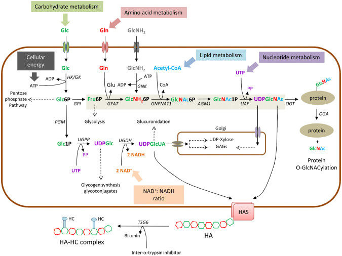
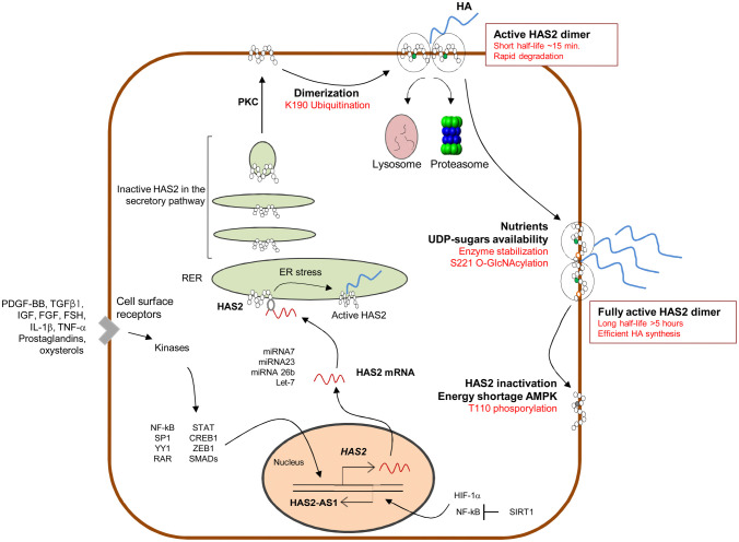
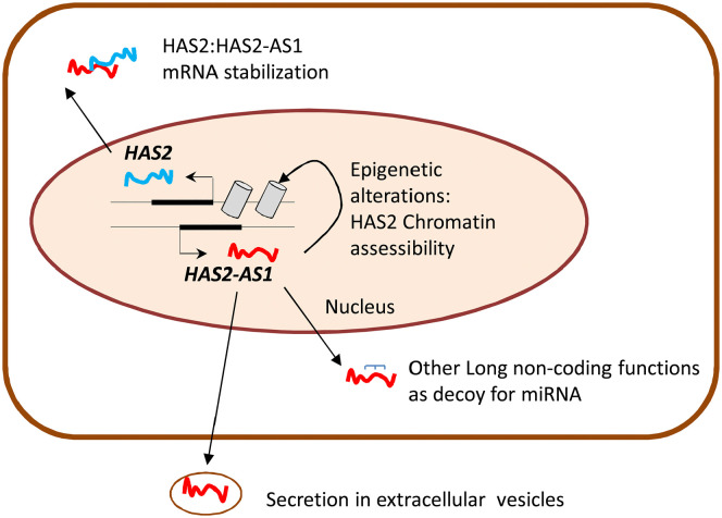
Hyaluronan (HA) is a linear glycosaminoglycan (GAG) of extracellular matrix (ECM) synthesized by three hyaluronan synthases (HASes) at the plasma membrane using uridine diphosphate (UDP)-glucuronic acid (UDP-GlcUA) and UDP-N-acetylglucosamine (UDP-GlcNAc) as substrates. The production of HA is mainly regulated by hyaluronan synthase 2 (HAS2), that can be controlled at different levels, from epigenetics to transcriptional and post-translational modifications. HA biosynthesis is an energy-consuming process and, along with HA catabolism, is strongly connected to the maintenance of metabolic homeostasis. The cytoplasmic pool of UDP-sugars is critical for HA synthesis. UDP-GlcNAc is an important nutrient sensor and serves as donor substrate for the O-GlcNAcylation of many cytosolic proteins, including HAS2. This post-translational modification stabilizes HAS2 in the membrane and increases HA production. Conversely, HAS2 can be phosphorylated by AMP activated protein kinase (AMPK), a master metabolic regulator activated by low ATP/AMP ratios, which inhibits HA secretion. Similarly, HAS2 expression and the deposition of HA within the pericellular coat are inhibited by sirtuin 1 (SIRT1), another important energetic sensor, confirming the tight connection between nutrients availability and HA metabolism.

Keywords: HAS2-AS1; UGDH; autophagy; beta glycosidases; cancer; cardiovascular diseases; extracellular matrix; glycosaminoglycan; hexosamine biosynthetic pathway; hyaluronidase; metabolic reprogramming; proteoglycan; ubiquitin.


Conflict of interest statement

Competing Interests: The author(s) declared no potential conflicts of interest with respect to the research, authorship, and/or publication of this article.

Figures

Figure 1.
Figure 1.
Schematic representation of the biosynthesis of UDP-GlcUA and UDP-GlcNAc (through the hexosamine biosynthetic pathway, shaded in light brown). Connections with the main biochemical pathways are indicated with dotted lines. Protein O-GlcNAcylation in the cytosol and GAGs synthesis in the Golgi are also shown. The mechanism of TSG6 adding HCs from IaI to form the HC-HA matrix is shown in the extracellular space. Abbreviation: Glc, glucose; Gln, glucosamine; GlcNH2, glucosamine; Glc6P, glucose-6-phosphate; Glc1P, glucose-1-phosphate; UDPGlc, UDP-glucose, UDPGlcUA, UDP-glucuronic acid; Fru6P, Fructose-6-phosphate, GlcNH26P, Glucosamine-6 phosphate; GlcNAc6P, N-acetyl-glucosamine-6-phosphate; GlcNAc1P, N-acetyl-glucosamine-1-phosphate; UDPGlcNAc, UDP-N-acetyl-glucosamine; Glu, glutamate; Acetyl-CoA, acetyl-coenzyme A; PP, pyrophosphate. HK; hexokinase; GK, glucokinase; GPI, glucose-6-phosphate isomerase; PGM, phospho glucomutase; UGPP, UDP-Glucose pyrophosphorylase; UGDH, UDP-Glucose dehydrogenase; GFAT, glutamine: fructose-6-phosphate amidotransferase; GNK, glucosamine kinase; GNPNAT1, GlcNH 2-6-phosphate N-acetyltransferase; AGM1, phospho-GlcNAc mutase; UAP, UDP-GlcNAc pyrophosphorylase; OGT, O-GlcNAc transferase; OGA; O-GlcNAcase.
Figure 2.
Figure 2.
Schematic representation of three main degradation pathways of HA through HYALs, HYBID and TMEM2. Connections with the main biochemical pathways are indicated with dotted lines. GlcNAc salvage shaded in light blue.
Figure 3.
Figure 3.
Schematic representation of HAS2 regulation. Several soluble molecules induce activation of transcription factors able to activate HAS2 transcription; HAS2 mRNA interacts with several miRNA altering stability and translation; HAS2 protein follows the secretory pathway to reach the plasma membrane; HAS2 ubiquitination leads to dimerization which represent the active HAS2 on the membrane. In this state, HAS2 is a very rapidly degraded enzyme; HAS2 O-GlcNAcylation strongly increases half-life of the enzyme in the membrane; HAS2 phosphorylation by AMPK blocks HAS2 activity. NF-kB and HIF-1α modulate HAS2-AS1 transcription; SIRT1 inhibits HAS2-AS1 that, in turn, reduces HAS2 transcription.
Figure 4.
Figure 4.
Schematic representation of cytoplasmic and nuclear functions of HAS2-AS1.

References

    1. Lu P, Takai K, Weaver VM, Werb Z. Extracellular Matrix degradation and remodeling in development and disease. Cold Spring Harb Perspect Biol. 2011;3:a005058. - PMC - PubMed
    1. Karamanos NK, Theocharis AD, Neill T, Iozzo RV. Matrix modeling and remodeling: a biological interplay regulating tissue homeostasis and diseases. Matrix Biol. 2019;75–76:1–11. - PMC - PubMed
    1. Sainio A, Järveläinen H. Extracellular matrix-cell interactions: focus on therapeutic applications. Cell Signal. 2020;66:109487. - PubMed
    1. Maquart FX, Pasco S, Ramont L, Monboisse J-C. An introduction to matrikines: extracellular matrix—derived peptides which regulate cell activity—implication in tumor invasion. Crit Rev Oncol/Hematol. 2004;49:199–202. - PubMed
    1. Wells JM, Gaggar A, Blalock JE. MMP generated matrikines. Matrix Biol. 2015;44–46:122–9. - PMC - PubMed

Publication types

MeSH terms

Substances