

Dissociable Networks of the Lateral/Medial Mammillary Body in the Human Brain
- PMID: 32625073
- PMCID: PMC7316159
- DOI: 10.3389/fnhum.2020.00228
Dissociable Networks of the Lateral/Medial Mammillary Body in the Human Brain
Abstract
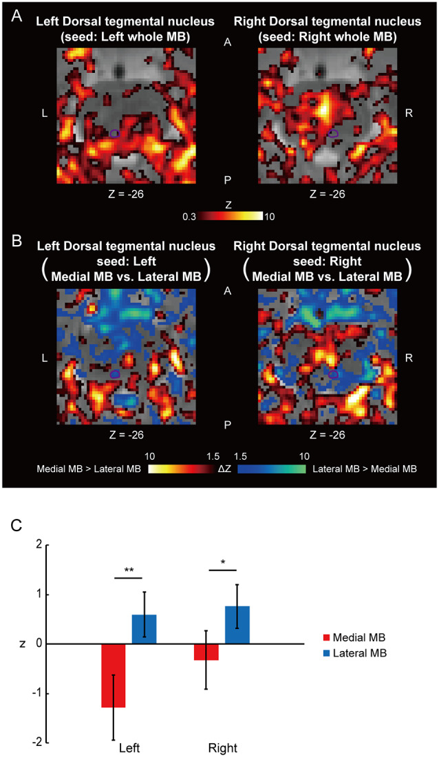
The mammillary body (MB) has been thought to implement mnemonic functions. Although recent animal studies have revealed dissociable roles of the lateral and medial parts of the MB, the dissociable roles of the lateral/medial MB in the human brain is still unclear. Functional connectivity using resting-state functional magnetic resonance imaging (fMRI) provides a unique opportunity to noninvasively inspect the intricate functional organization of the human MB with a high degree of spatial resolution. The present study divided the human MB into lateral and medial parts and examined their functional connectivity with the hippocampal formation, tegmental nuclei, and anterior thalamus. The subiculum of the hippocampal formation was more strongly connected with the medial part than with the lateral part of the MB, whereas the pre/parasubiculum was more strongly connected with the lateral part than with the medial part of the MB. The dorsal tegmental nucleus was connected more strongly with the lateral part of the MB, whereas the ventral tegmental nucleus showed an opposite pattern. The anterior thalamus was connected more strongly with the medial part of the MB. These results confirm the extant animal literature on the lateral/medial MB and provide evidence on the parallel but dissociable systems involving the MB that ascribe mnemonic and spatial-navigation functions to the medial and lateral MBs, respectively.
Keywords: fornix; hippocampus; mammillotegmental tract; resting-state functional connectivity; tegmentum.
Copyright © 2020 Tanaka, Osada, Ogawa, Kamagata, Aoki and Konishi.
Figures


Similar articles
-
An autoradiographic study of the organization of the efferent connections of the hippocampal formation in the rat.J Comp Neurol. 1977 Mar 1;172(1):49-84. doi: 10.1002/cne.901720104. J Comp Neurol. 1977. PMID: 65364
-
Retrograde double-labeling study of the mammillothalamic and the mammillotegmental projections in the rat.J Comp Neurol. 1989 Jun 1;284(1):1-11. doi: 10.1002/cne.902840102. J Comp Neurol. 1989. PMID: 2502564
-
The connections of the septal region in the rat.J Comp Neurol. 1979 Aug 15;186(4):621-55. doi: 10.1002/cne.901860408. J Comp Neurol. 1979. PMID: 15116692
-
Mamillary body in the rat: topography and synaptology of projections from the subicular complex, prefrontal cortex, and midbrain tegmentum.J Comp Neurol. 1989 Aug 15;286(3):311-36. doi: 10.1002/cne.902860303. J Comp Neurol. 1989. PMID: 2504784
-
Neurochemistry of the mammillary body.Brain Struct Funct. 2023 Jul;228(6):1379-1398. doi: 10.1007/s00429-023-02673-4. Epub 2023 Jun 28. Brain Struct Funct. 2023. PMID: 37378855 Free PMC article. Review.
Cited by
-
Multiple insular-prefrontal pathways underlie perception to execution during response inhibition in humans.Nat Commun. 2024 Dec 3;15(1):10380. doi: 10.1038/s41467-024-54564-9. Nat Commun. 2024. PMID: 39627197 Free PMC article.
-
Performance ramifications of abnormal functional connectivity of ventral posterior lateral thalamus with cerebellum in abstinent individuals with Alcohol Use Disorder.Drug Alcohol Depend. 2021 Mar 1;220:108509. doi: 10.1016/j.drugalcdep.2021.108509. Epub 2021 Jan 8. Drug Alcohol Depend. 2021. PMID: 33453503 Free PMC article.
-
Stereotaxic Coordinates of Human Hypothalamic Nuclei Used for Region of Interest Analyses in Functional Magnetic Resonance Imaging.Juntendo Iji Zasshi. 2024 Apr 18;70(2):129-131. doi: 10.14789/jmj.JMJ24-0009-P. eCollection 2024. Juntendo Iji Zasshi. 2024. PMID: 39430209 Free PMC article.
-
Functional Organization for Response Inhibition in the Right Inferior Frontal Cortex of Individual Human Brains.Cereb Cortex. 2020 Nov 3;30(12):6325-6335. doi: 10.1093/cercor/bhaa188. Cereb Cortex. 2020. PMID: 32666077 Free PMC article.
-
Compromised mammillary body connectivity and psychotic symptoms in mice with di- and mesencephalic ablation of ST8SIA2.Transl Psychiatry. 2022 Feb 3;12(1):51. doi: 10.1038/s41398-022-01816-1. Transl Psychiatry. 2022. PMID: 35115485 Free PMC article.
References
LinkOut - more resources
Full Text Sources
Miscellaneous