. 2020 Jun 17:11:922.
doi: 10.3389/fphar.2020.00922. eCollection 2020.

Synthesis and Biological Evaluation of the Anti-Melanogenesis Effect of Coumaric and Caffeic Acid-Conjugated Peptides in Human Melanocytes

Affiliations

Synthesis and Biological Evaluation of the Anti-Melanogenesis Effect of Coumaric and Caffeic Acid-Conjugated Peptides in Human Melanocytes

Kyeong-Yong Park et al. Front Pharmacol. .

Abstract

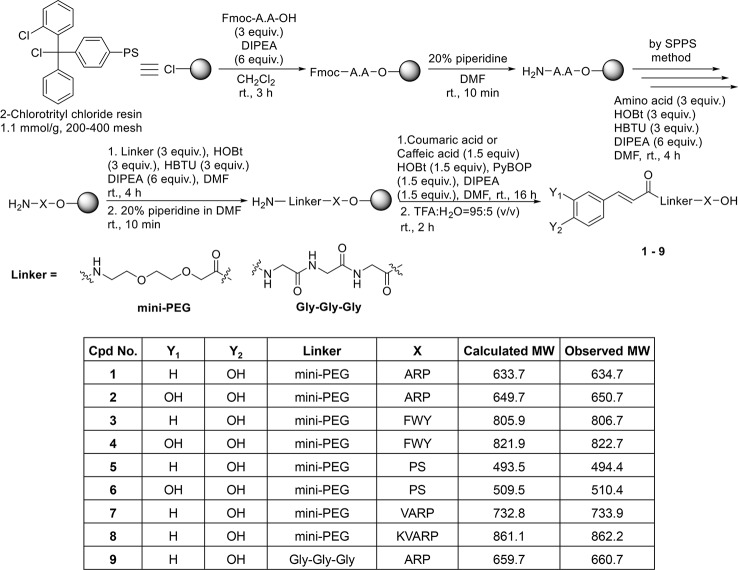
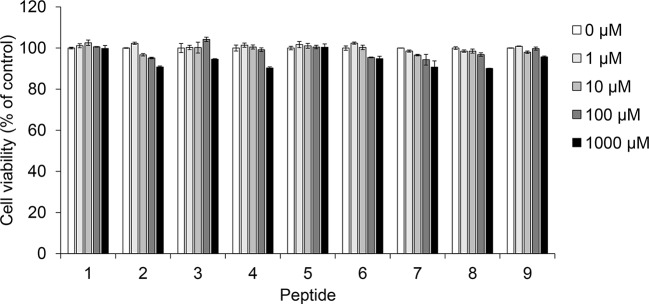
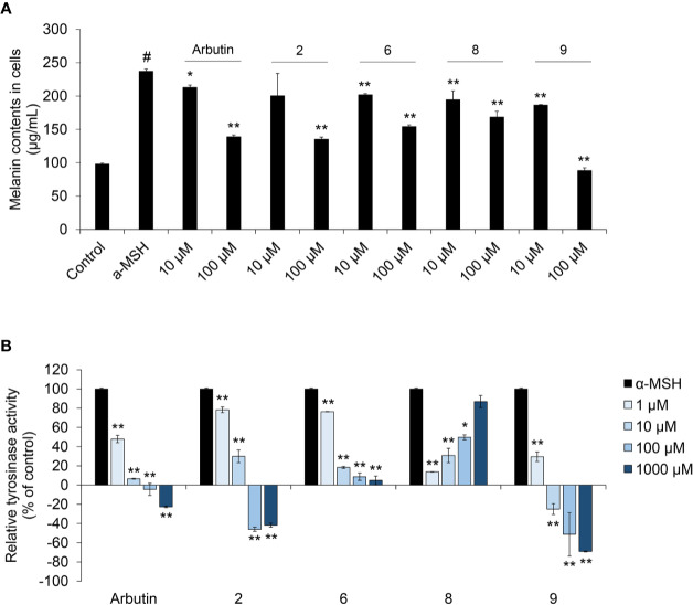
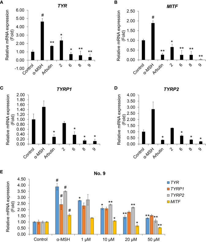
Excessive pigmentation and reduced elasticity are the major skin problems that dermatologists and cosmetologists address. Compounds that inhibit melanin production might contribute to improving skin problems. In this study, we investigated whether coumaric acid- and caffeic acid-conjugated peptides might affect alpha-melanocyte stimulating hormone-induced melanin production, tyrosinase activity, and melanin synthesis-related gene expression in SK-MEL-2 human melanoma cells. Coumaric acid and caffeic acid showed no significant cytotoxicity, and they inhibited melanin production. In addition, coumaric acid- and caffeic acid-conjugated peptides suppressed tyrosinase activity more than arbutin, a known tyrosinase inhibitor. Quantitative real-time PCR (qRT-PCR) results also showed that both peptides inhibited the expression of melanin synthesis-related genes, TYR, TYRP1, TYRP2, and MITF. In particular, among the nine conjugated peptides tested, caffeic acid linked to a Gly-Gly-Gly linker and conjugated to the tripeptide, ARP, showed the greatest inhibition of gene expression in the qRT-PCR analysis. These results suggested that the inhibition of melanin exerted by coumaric acid- and caffeic acid-conjugated peptides might provide important information for the development of pigmentation-related skin diseases and cosmetic products.

Keywords: SK-MEL-2; coumaric acid; melanin; tyrosinase; α-MSH.


Figures

Figure 1
Figure 1
Solid phase synthesis of coumaric acid- and caffeic acid-peptide conjugates.
Figure 2
Figure 2
The effect of coumaric acid- and caffeic acid-peptide conjugates on the viability of SK-MEL-2 cells. SK-MEL-2 cells were treated with coumaric acid- or caffeic acid-conjugated peptides (19) for 72 h. Cell viability is expressed as the percent of control (control: 0 μM). Experiments were performed in triplicate. Statistical significance is represented in Supplementary Table S2.
Figure 3
Figure 3
The effect of coumaric acid- and caffeic acid-peptide conjugates on α-MSH –induced melanin synthesis and tyrosinase activity in SK-MEL-2 cells. SK-MEL-2 cells were treated with α-MSH (200 nM) alone or with the indicated concentrations of peptides for 72 h. (A) Melanin content in cells (μg/ml). (B) Relative tyrosinase inhibition is expressed as a percentage of control (control: α-MSH only). Data represent the mean ± SD of experiments performed in triplicate. #p < 0.01 versus control, *p < 0.05, **p < 0.01 versus treatment with α-MSH alone.
Figure 4
Figure 4
The effects of coumaric acid- and caffeic acid-peptide conjugates on the expression of melanogenesis-related genes. (A–D) Relative gene expression, expressed as the fold change in mRNA levels compared to control (untreated) in SK-MEL-2 cells. Cells were treated with α-MSH (200 nM), without or with 100 μM of coumaric acid- or caffeic acid-conjugated peptides (2, 6, 8, 9) or arbutin for 48 h. (E) Relative gene expression, expressed as the fold change in mRNA levels in SK-MEL-2 cells, compared to control (untreated) cells. Cells were treated with α-MSH (200 nM) without or with different concentrations of compound 9 (1–50 μM) for 48 h. GAPDH served as the internal control. Data represent the mean ± SD of experiments performed in triplicate. #p < 0.01 versus control, *p < 0.05, **p < 0.01 versus treatment with α-MSH only.

Similar articles

Cited by

References

    1. Aoki H., Moro O. (2012). Involvement of microphthalmia-associated transcription factor (MITF) in expression of human melanocortin-1 receptor (MC1R). Life Sci. 71, 2171–2179. 10.1016/S0024-3205(02)01996-3 - DOI - PubMed
    1. Bertolotto C., Buscà R., Abbe P., Bille K., Aberdam E., Ortonne J. P., et al. (1998). Different cis-acting elements are involved in the regulation of TRP1 and TRP2 promoter activities by cyclic AMP: pivotal role of M boxes (GTCATGTGCT) and of microphthalmia. Mol. Cell Biol. 18, 694–702. 10.1128/MCB.18.2.694 - DOI - PMC - PubMed
    1. Boo Y. C. (2019). p-Coumaric Acid as An Active Ingredient in Cosmetics: A Review Focusing on its Antimelanogenic Effects. Antioxidants (Basel) 8, E275. 10.3390/antiox8080275 - DOI - PMC - PubMed
    1. Cabanes J., Chazarra S., Garcia-Carmona F. (1994). Kojic acid, a cosmetic skin whitening agent, is a slow-binding inhibitor of catecholase activity of tyrosinase. J. Pharm. Pharmacol. 46, 982–985. 10.1111/j.2042-7158.1994.tb03253.x - DOI - PubMed
    1. Chakraborty A., Slominski A., Ermak G., Hwang J., Pawelek J. (1995). Ultraviolet B and melanocyte-stimulating hormone (MSH) stimulate mRNA production for alpha MSH receptors and proopiomelanocortin-derived peptides in mouse melanoma cells and transformed keratinocytes. J. Invest. Dermatol. 105, 655–659. 10.1111/1523-1747.ep12324134 - DOI - PubMed