. 2020 Jun 18:11:1222.
doi: 10.3389/fmicb.2020.01222. eCollection 2020.

Surface Protein Dispersin of Enteroaggregative Escherichia coli Binds Plasminogen That Is Converted Into Active Plasmin

Affiliations

Surface Protein Dispersin of Enteroaggregative Escherichia coli Binds Plasminogen That Is Converted Into Active Plasmin

Claudia T P Moraes et al. Front Microbiol. .

Abstract

Dispersin is a 10.2 kDa-immunogenic protein secreted by enteroaggregative Escherichia coli (EAEC). In the prototypical EAEC strain 042, dispersin is non-covalently bound to the outer membrane, assisting dispersion across the intestinal mucosa by overcoming electrostatic attraction between the AAF/II fimbriae and the bacterial surface. Also, dispersin facilitates penetration of the intestinal mucus layer. Initially characterized in EAEC, dispersin has been detected in other E. coli pathotypes, including those isolated from extraintestinal sites. In this study we investigated the binding capacity of purified dispersin to extracellular matrix (ECM), since dispersin is exposed on the bacterial surface and is involved in intestinal colonization. Binding to plasminogen was also investigated due to the presence of conserved carboxy-terminal lysine residues in dispersin sequences, which are involved in plasminogen binding in several bacterial proteins. Moreover, some E. coli components can interact with this host protease, as well as with tissue plasminogen activator, leading to plasmin production. Recombinant dispersin was produced and used in binding assays with ECM molecules and coagulation cascade compounds. Purified dispersin bound specifically to laminin and plasminogen. Interaction with plasminogen occurred in a dose-dependent and saturable manner. In the presence of plasminogen activator, bound plasminogen was converted into plasmin, its active form, leading to fibrinogen and vitronectin cleavage. A collection of E. coli strains isolated from human bacteremia was screened for the presence of aap, the dispersin-encoding gene. Eight aap-positive strains were detected and dispersin production could be observed in four of them. Our data describe new attributes for dispersin and points out to possible roles in mechanisms of tissue adhesion and dissemination, considering the binding capacity to laminin, and the generation of dispersin-bound plasmin(ogen), which may facilitate E. coli spread from the colonization site to other tissues and organs. The cleavage of fibrinogen in the bloodstream, may also contribute to the pathogenesis of sepsis caused by dispersin-producing E. coli.

Keywords: EAEC; ECM; dispersin; plasmin; plasminogen.


Figures

FIGURE 1
FIGURE 1
Purified recombinant dispersin. (A) SDS-PAGE (15%) of purified recombinant dispersin (∼10.2 kDa) after Coomassie Brilliant Blue staining. Kaleidoscope Protein Standards (Bio-Rad, Hercules, CA, United States) was used as protein size marker. (B) Immunoblotting of recombinant dispersin using anti-dispersin serum produced in rabbit (1:100). PageRuler Prestained Protein Ladder (Thermo Fisher Scientific, Waltham, MA, United States) was used as protein size marker.
FIGURE 2
FIGURE 2
Binding of recombinant dispersin to extracellular matrix (ECM) components and coagulation cascade molecules. ELISA plate wells were coated with 1 μg of ECM components (CF, cellular fibronectin; LAM, laminin; COL I, collagen I; and COL IV, collagen IV) and plasma proteins (PP) (PF, plasma fibronectin; FB, fibrinogen; and PLG, plasminogen). Bovine serum albumin (BSA) was included as negative control. One microgram of recombinant dispersin was added per well. Bound proteins were detected using specific rabbit antiserum to the recombinant protein, followed by peroxidase-conjugated antibodies. Bars represent the mean absorbance value at 492 nm ± standard deviation of three independent experiments, performed in triplicates. For these analyses, Student’s t-test was used (***P = 0.0002; ****P < 0.0001).
FIGURE 3
FIGURE 3
Dose-response binding of recombinant dispersin to purified human plasminogen by surface plasmon resonance (SPR). Interaction of dispersin with plasminogen in the BIAcore T100 system. Dispersin at different concentrations was injected over immobilized plasminogen at a flow rate of 30 μL/min. RU, response units.
FIGURE 4
FIGURE 4
Plasminogen bound to dispersin can be converted in active plasmin. Recombinant dispersin, MntC or BSA (1 μg) were immobilized onto microplate wells and then incubated with plasminogen. After that, uPA (3 U) and the chromogenic substrate for plasmin D-valyl-leucyl-lysine-ρ-nitroanilide dihydrochloride (25 μg/well) were added. Bars represent the mean absorbance value at 492 nm ± standard deviation of three independent experiments, performed in triplicates. For this analysis, Student’s t-test was used (**p = 0.0012; ****p = 0.0001). P, dispersin, MntC or BSA; PLG, plasminogen; uPa, human urokinase plasminogen activator; Sub, chromogenic substrate; PAI-1, plasminogen activator inhibitor 1.
FIGURE 5
FIGURE 5
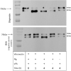
Degradation of human fibrinogen (Fbg) by plasmin(ogen) bound to immobilized dispersin. Recombinant dispersin (10 μg/mL), purified MntC (positive control) and BSA (negative control) were immobilized onto microplate wells and incubated with 10 μg/mL of plasminogen (Plg). After washing, human fibrinogen (10 μg) and plasminogen activator uPA (3 U) were added. After incubation during 1 or 4 h, proteins were separated by SDS-PAGE and transferred to a polyvinylidene difluoride membrane for immunodetection with anti-fibrinogen antibodies (1:1,000) (Cloud-Clone, Katy, TX, United States). Asterisks show fibrinogen cleavage fragments: a band between β and γ chains of fibrinogen was observed in presence of dispersin and MntC, but was not observed in the presence of BSA (negative control), after 4 h of incubation. Kaleidoscope Protein Standards (Bio-Rad, Hercules, CA, United States) was used as protein size marker.
FIGURE 6
FIGURE 6
Degradation of human vitronectin by plasmin(ogen) bound to immobilized dispersin. Recombinant dispersin (10 μg/mL) and BSA (negative control) were immobilized onto microplate wells and incubated with 10 μg/mL of plasminogen (Plg). After washing, human vitronectin (1 μg) and plasminogen activator uPA (3 U) were added. After incubation during 1 or 4 h, proteins were separated by SDS-PAGE and transferred to a polyvinylidene difluoride membrane for immunodection with anti-vitronectin antibodies (1:5,000) (Complement Technology, Tyler, TX, United States). Asterisk shows vitronectin degradation: the isoform of 75 kDa was completely degraded in the presence of dispersin, but not in the presence of BSA (negative control), after 4 h of incubation. Kaleidoscope Protein Standards (Bio-Rad, Hercules, CA, United States) was used as protein size marker.
FIGURE 7
FIGURE 7
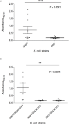
Detection of dispersin by ELISA in E. coli strains isolated from blood of human patients. Bacterial pellets of overnight cultures in LB broth were used for coating of ELISA plates, followed by incubation with anti-dispersin polyclonal serum (1:200) produced in rabbit. After washing, horseradish peroxidase-conjugated goat anti-rabbit IgG (Sigma-Aldrich) was added (1:1,000). Blank wells were performed in absence of anti-dispersin sera. EAEC 17-2 and 042 were considered as the positive controls. Data represent the mean absorbance value at 492 nm ± the standard deviation of three independent experiments, performed in triplicates. (A) Groups aap-positive and aap-negative were analyzed by non-parametric Student’s t-test (****P ≤ 0.0001) with 95% confidence interval: –0.02416 to 1.086. (B) Differences of dispersin production between aap-positive strains were analyzed by ANOVA test (**P = 0.0074) with 95% confidence intervals: aap+/dispersin+: 0.1963–1.932; aap+/dispersin–: 0.153–0.2157; aap–/dispersin–: 0.1171–0.2311.
FIGURE 8
FIGURE 8
Schematic model of the proposed role for dispersin in bacterial dissemination. (A) Dispersin-producing E. coli reaches the intestinal or urinary tracts adhering to those epithelia using diverse mechanisms, including binding to laminin; (B) Those bacteria presenting invasiveness capacity may penetrate the epithelia getting in contact with connecting tissues, microvessels and the bloodstream; (C) In these sites, dispersin exposed on the bacterial surface can bind to Plasminogen; (D) Plasminogen bound to Dispersin can be activated into Plasmin in the presence of plasminogen activators (uPa and tPa); (E) Active Plasmin can act as a potent protease promoting degradation of physiologic subtracts such as fibrin clots and/or extracellular matrix proteins; (F) These two effects lead to bacterial dissemination. Plg, plasminogen; Pla, plasmin; uPa, urokinase plasminogen activator; tPa, tissue plasminogen activator; ECM, extracellular matrix proteins.

References

    1. Abe C. M., Knutton S., Pedroso M. Z., Freymüller E., Gomes T. A. (2001). An enteroaggregative Escherichia coli strain of serotype O111:H12 damages and invades cultured T84 cells and human colonic mucosa. FEMS Microbiol. Lett. 203 199–205. 10.1111/j.1574-6968.2001.tb10841.x - DOI - PubMed
    1. Abe C. M., Salvador F. A., Falsetti I. N., Vieira M. A. M., Blanco J., Blanco J. E., et al. (2008). Uropathogenic Escherichia coli (UPEC) strains may carry virulence properties of diarrhoeagenic E. coli. FEMS Immunol. Med. Microbiol. 52 397–406. 10.1111/j.1574-695X.2008.00388.x - DOI - PubMed
    1. Abreu A. G., Barbosa A. S. (2017). How Escherichia coli circumvent complement-mediated killing. Front. Immunol. 8:452 10.3389/fimmu.2017.00452 - DOI - PMC - PubMed
    1. Altschul S. F., Madden T. L., Schäffer A. A., Zhang J., Zhang Z., Miller W., et al. (1997). Gapped BLAST and PSI-BLAST: new generation of protein database search programs. Nucleic Acids Res. 25 3389–3402. 10.1093/nar/25.17.3389 - DOI - PMC - PubMed
    1. Andrade F. B., Gomes T. A. T., Elias W. P. (2014). A sensitive and specific molecular tool for detection of both typical and atypical enteroaggregative Escherichia coli. J. Microbiol. Methods 106 16–18. 10.1016/j.mimet.2014.07.030 - DOI - PubMed