Review
. 2020 Jun 17:11:1234.
doi: 10.3389/fimmu.2020.01234. eCollection 2020.

Emerging Roles of Mast Cells in the Regulation of Lymphatic Immuno-Physiology

Affiliations
Review

Emerging Roles of Mast Cells in the Regulation of Lymphatic Immuno-Physiology

Sarit Pal et al. Front Immunol. .

Abstract

Mast cells (MCs) are abundant in almost all vascularized tissues. Furthermore, their anatomical proximity to lymphatic vessels and their ability to synthesize, store and release a large array of inflammatory and vasoactive mediators emphasize their significance in the regulation of the lymphatic vascular functions. As a major secretory cell of the innate immune system, MCs maintain their steady-state granule release under normal physiological conditions; however, the inflammatory response potentiates their ability to synthesize and secrete these mediators. Activation of MCs in response to inflammatory signals can trigger adaptive immune responses by dendritic cell-directed T cell activation. In addition, through the secretion of various mediators, cytokines and growth factors, MCs not only facilitate interaction and migration of immune cells, but also influence lymphatic permeability, contractility, and vascular remodeling as well as immune cell trafficking through the lymphatic vessels. In summary, the consequences of these events directly affect the lymphatic niche, influencing inflammation at multiple levels. In this review, we have summarized the recent advancements in our understanding of the MC biology in the context of the lymphatic vascular system. We have further highlighted the MC-lymphatic interaction axis from the standpoint of the tumor microenvironment.

Keywords: cancer; immune response; lymphatic system; lymphatic vessels; mast cells.


Figures

Figure 1
Figure 1
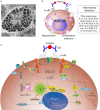
Overview of MC activation and degranulation mechanisms. (A) A transmission electron microscope image of an activated MC showing multiple secretory granules inside the cell. Adapted from Grujic et al. (25) and reproduced with written permission from the publisher. Copyright 2013, the American Association of Immunologists, Inc. (B) A schematic of a MC showing Immunoglobulin E (IgE)-mediated interaction with allergen and secretion of different inflammatory mediators. (C). Aggregation of the IgE Receptor (FcεRI) by multivalent antigen induces activation of tyrosine-protein kinase Lyn (Lyn), the Src kinase that phosphorylates immunoreceptor tyrosine-based activation motifs (ITAMs) of FcεRI β and γ subunits, followed by the association of the tyrosine-protein kinase Syk with the FcεRI via Syk-Src Homology domain 2 (SH2) within phosphorylated ITAMs. This clustering leads to activation of tyrosine-protein kinase Fyn that phosphorylates the adaptor growth factor receptor-bound protein 2 (Grb2). Activation of phospholipase C gamma 1 (PLC-γ1) results in the hydrolysis of phosphatidylinositol-4,5-bisphosphate (PIP2) into inositol 1, 4, 5-triphosphate (IP3) and diacylglycerol (DAG). IP3 production leads to increased intracellular free calcium (Ca2+) concentration, whereas DAG can activate both protein kinase C-θ (PKC-θ) and Ras. Tyrosine phosphorylated SLP76 also associates with the Rho-family guanine nucleotide exchange factor (GEF) Vav1 and the adaptor protein, Nck. Vav1 activates Rac and cell division control protein 42 (Cdc42), which initiate actin cytoskeletal rearrangement via activation of Wiskott-Aldrich syndrome protein (WASP). Cytoskeletal rearrangement is required for cell migration and microtubule-dependent degranulation of MCs.
Figure 2
Figure 2
Lymphatic system architecture along with the lymphatic tissue niche. (1) Initial lymphatics (lymphatic capillaries) (2) Pre-collector LVs; (3) Perilymphatic antigen-presenting cells; (4) Perilymphatic MCs; (5) Collecting LVs; (6) Lymphatic valve in collecting LVs; (7) Degranulating MCs in response to inflammation; (8) Increased permeability of LVs in response to inflammation; (9) LECs; (10) Lymphatic muscle cells; and (11) Inflammation-induced MC-DC immune synapse formation in perilymphatic tissues. dLN, Draining lymph node.
Figure 3
Figure 3
Mast cells in the tumor microenvironment. (A) MCs secrete different cytokines/inflammatory mediators into the tumor microenvironment. These cytokines/inflammatory mediators modulate cellular and acellular components of the tumor. The cellular components include cancer cells, cancer-associated fibroblasts, and different types of immune cells, while the acellular component is mainly the extracellular matrix (ECM). (B) MCs also modulate angiogenesis and lymphangiogenesis in the tumor. Tumor necrosis factor-alpha (TNFα) released by MCs induces migration of DCs into the draining lymph nodes where T cell activation takes place. MC-derived proteases induce modification of the ECM, which alters the microarchitecture leading to metastasis.

References

    1. Theoharides TC, Valent P, Akin C. Mast cells mastocytosis, and related disorders. N Engl J Med. (2015) 373:163–72. 10.1056/NEJMra1409760 - DOI - PubMed
    1. Ngkelo A, Richart A, Kirk JA, Bonnin P, Vilar J, Lemitre M, et al. . Mast cells regulate myofilament calcium sensitization and heart function after myocardial infarction. J Exp Med. (2016) 213:1353–74. 10.1084/jem.20160081 - DOI - PMC - PubMed
    1. Miller HR, Pemberton AD. Tissue-specific expression of mast cell granule serine proteinases and their role in inflammation in the lung and gut. Immunology. (2002) 105:375–90. 10.1046/j.1365-2567.2002.01375.x - DOI - PMC - PubMed
    1. Ehara T, Shigematsu H. Mast cells in the kidney. Nephrology. (2003) 8:130–8. 10.1046/j.1440-1797.2003.00153.x - DOI - PubMed
    1. Bischoff SC, Kramer S. Human mast cells, bacteria, and intestinal immunity. Immunol Rev. (2007) 217:329–37. 10.1111/j.1600-065X.2007.00523.x - DOI - PubMed

Publication types