. 2020 Aug;22(2):1558-1566.
doi: 10.3892/mmr.2020.11211. Epub 2020 Jun 3.

Siva‑1 regulates multidrug resistance of gastric cancer by targeting MDR1 and MRP1 via the NF‑κB pathway

Affiliations

Siva‑1 regulates multidrug resistance of gastric cancer by targeting MDR1 and MRP1 via the NF‑κB pathway

Fan-Biao Kong et al. Mol Med Rep. 2020 Aug.

Abstract

Siva‑1 is a well‑known anti‑apoptosis protein that serves a role in multiple types of cancer cells. However, whether Siva‑1 affects multidrug resistance via the NF‑κB pathway in gastric cancer is currently unknown. The present study aimed to determine the possible involvement of Siva‑1 in gastric cancer anticancer drug resistance in vitro. A vincristine (VCR)‑resistant KATO III/VCR gastric cancer cell line with stable Siva‑1 overexpression was established. The protein expression levels of Siva‑1, NF‑κB, multidrug resistance 1 (MDR1) and multidrug resistance protein 1 (MRP1) were detected via western blotting. The effect of Siva‑1 overexpression on anticancer drug resistance was assessed by measuring the 50% inhibitory concentration of KATO III/VCR cells to VCR, 5‑fluorouracil and doxorubicin. The rate of doxorubicin efflux and apoptosis were detected by flow cytometry. Additionally, colony formation, wound healing and Transwell assays were used to detect the proliferation, migration and invasion of cells, respectively. The results of the current study revealed that the Siva‑1‑overexpressed KATO III/VCR gastric cancer cells exhibited a significantly decreased sensitivity to VCR, 5‑fluorouracil and doxorubicin. The results of flow cytometry revealed that the percentage of apoptotic cells decreased following overexpression of Siva‑1. The colony formation assay demonstrated that cell growth and proliferation were significantly promoted by Siva‑1 overexpression. Additionally, Siva‑1 overexpression increased the migration and invasion of KATO III/VCR cells in vitro. Western blot analysis determined that Siva‑1 overexpression increased NF‑κB, MDR1 and MRP1 levels. The current study demonstrated that overexpression of Siva‑1, which functions as a regulator of MDR1 and MRP1 gene expression in gastric cancer cells via promotion of NF‑κB expression, inhibited the sensitivity of gastric cancer cells to certain chemotherapies. These data provided novel insight into the molecular mechanisms of gastric cancer, and may be of significance for the clinical diagnosis and therapy of patients with gastric cancer.

Keywords: Siva-1; multidrug resistance; gastric cancer.


Figures

Figure 1.
Figure 1.
Recombinant pGV358-GFP-SIVA-1 plasmid was transfected into 293T cells to determine lentivirus titers using the end-point dilution method, which involved counting the number of infected green cells under fluorescence microscopy (magnification, ×100). The viral dilution factor was 1:1,000. LV, lentivirus; NC, negative control; MOI, multiplicity of infection. (A) In light microscope; (B) in fluorescence microscopy.
Figure 2.
Figure 2.
Siva-1, NF-κB, MDR1 and MRP1 protein expression is determined via western blotting. MDR1 and MRP1 protein levels were increased following Siva-1 overexpression and NF-κB was active following its rapid translocation into the nucleus after the same treatment. (A) Western blot analysis and (B) subsequent semi-quantification of Siva-1, MDR1 and MRP1 protein levels in the three groups. (C) Western blot analysis and (D) subsequent semi-quantification of NF-κB protein levels in the three groups. Expression was normalized to that of GAPDH or Lamin B1, and presented as the mean ± standard deviation. *P<0.05 vs. group 2 and 3. 1, KATO III/VCR + LV-Siva-1; 2, KATO III/VCR + LV-NC; 3, KATO III/VCR. MDR1, multidrug resistance 1; MRP1, multidrug resistance protein 1; VCR, vincristine; LV, lentivirus; NC, negative control.
Figure 3.
Figure 3.
IC50 values for anticancer drugs applied to KATO III/VCR cells were evaluated using a MTT assay. Data were presented as the mean ± standard deviation of four independent experiments. *P<0.05 vs. group 2 and 3. 1, KATO III/VCR + LV-Siva-1; 2, KATO III/VCR + LV-NC; 3, KATO III/VCR. VCR, vincristine; LV, lentivirus; NC, negative control; IC50, 50% inhibitory concentration.
Figure 4.
Figure 4.
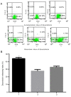
Pump rate of doxorubicin in KATO III/VCR cells after Siva-1 gene transfection. (A) Pump rate was analyzed by flow cytometry and (B) plotted. Data are presented as the mean ± standard deviation. *P<0.05 vs. group 2 and 3. 1, KATO III/VCR + LV-Siva-1; 2, KATO III/VCR + LV-NC; 3, KATO III/VCR. VCR, vincristine; LV, lentivirus; NC, negative control; DOX, doxorubicin.
Figure 5.
Figure 5.
Effect of Siva-1 overexpression on KATO III/VCR cell growth. (A and B) Apoptotic rate in Siva-1 overexpressed-KATO III/VCR cells was analyzed by flow cytometry. (C and D) KATO III/VCR + LV-Siva-1 cells, KATO III/VCR + LV-NC cells and KATO III/VCR cells were plated in 6-well plates at a density of 200 cells/well, after which colony growth was observed under an optical microscope following 14 days (magnification, ×40). The surviving fraction of cells (visible colonies) was stained with gentian violet and counted manually. Data are presented as the mean ± standard deviation from 3 independent experiments. *P<0.05 vs. group 2 and 3. 1, KATO III/VCR + LV-Siva-1; 2, KATO III/VCR + LV-NC; 3, KATO III/VCR; VCR, vincristine; LV, lentivirus; NC, negative control; 7-AAD, 7-amino-actinomyosin D.
Figure 6.
Figure 6.
Siva-1 overexpression increases KATO III/VCR cell migration and invasion. (A and B) KATO III/VCR + LV-Siva-1 cells, KATO III/VCR + LV-NC cells and KATO III/VCR cells were cultured to confluence on 6-well plates, after which a central linear wound was created with a 200-µl sterile pipette tip. The wound was imaged over a 4-day interval (magnification, ×40). (C and D) KATO III/VCR + LV-Siva-1 cells, KATO III/VCR + LV-NC cells and KATO III/VCR cells were loaded into the upper chambers of a Matrigel-coated Transwell plate. Filtrated cells on the undersurface of the polycarbonate membranes were stained and counted under an optical microscope after 48 h (magnification, ×200). *P<0.05 vs. group 2 and 3. 1, KATO III/VCR + LV-Siva-1; 2, KATO III/VCR + LV-NC; 3, KATO III/VCR. VCR, vincristine; LV, lentivirus; NC, negative control.

References

    1. Emmings E, Mullany S, Chang Z, Landen CN, Jr, Linder S, Bazzaro M. Targeting mitochondria for treatment of chemoresistant ovarian cancer. Int J Mol Sci. 2019;20:229. doi: 10.3390/ijms20010229. - DOI - PMC - PubMed
    1. Kongsema M, Wongkhieo S, Khongkow M, Lam EW, Boonnoy P, Vongsangnak W, Wong Ekkabut J. Molecular mechanism of Forkhead box M1 inhibition by thiostrepton in breast cancer cells. Oncol Rep. 2019;42:953–962. - PMC - PubMed
    1. Zeng L, Liao Q, Zou Z, Wen Y, Wang J, Liu C, He Q, Weng N, Zeng J, Tang H, et al. Long non-coding RNA XLOC_006753 promotes the development of multidrug resistance in gastric cancer cells through the PI3K/AKT/mTOR signaling pathway. Cell Physiol Biochem. 2018;51:1221–1236. doi: 10.1159/000495499. - DOI - PubMed
    1. Chen S, Wu J, Jiao K, Wu Q, Ma J, Chen D, Kang J, Zhao G, Shi Y, Fan D, et al. MicroRNA-495-3p inhibits multidrug resistance by modulating autophagy through GRP78/mTOR axis in gastric cancer. Cell Death Dis. 2018;9:1070. doi: 10.1038/s41419-018-0950-x. - DOI - PMC - PubMed
    1. Kozovska Z, Gabrisova V, Kucerova L. Colon cancer: cancer stem cells markers, drug resistance and treatment. Biomed Pharmacother. 2014;68:911–916. doi: 10.1016/j.biopha.2014.10.019. - DOI - PubMed

MeSH terms

Substances