

Primary open angle glaucoma genetics: The common variants and their clinical associations (Review)
- PMID: 32626970
- PMCID: PMC7339808
- DOI: 10.3892/mmr.2020.11215
Primary open angle glaucoma genetics: The common variants and their clinical associations (Review)
Abstract
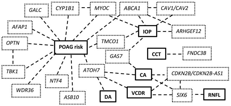
Glaucoma is a group of progressive optic neuropathies that have in common characteristic optic nerve head changes, loss of retinal ganglion cells and visual field defects. Among the large family of glaucomas, primary open‑angle glaucoma (POAG) is the most common type, a complex and heterogeneous disorder with environmental and genetic factors contributing to its pathogenesis. Approximately 5% of POAG is currently attributed to single‑gene or Mendelian forms of glaucoma. Genetic linkage analysis and genome‑wide association studies have identified various genomic loci, paving the path to understanding the pathogenesis of this enigmatic, blinding disease. In this review we summarize the most common variants reported thus far and their possible clinical correlations.
Keywords: glaucoma; primary open-angle glaucoma; genome-wide association studies; endophenotype; linkage; genetics.
Figures
References
-
- Gordon MO, Beiser JA, Brandt JD, Heuer DK, Higginbotham EJ, Johnson CA, Keltner JL, Miller JP, Parrish RK, II, Wilson MR, et al. The Ocular Hypertension Treatment Study: Baseline factors that predict the onset of primary open-angle glaucoma. Arch Ophthalmol. 2002;120:714–720, discussion 829–830. doi: 10.1001/archopht.120.6.714. - DOI - PubMed
-
- Fan BJ, Leung YF, Wang N, Lam SC, Liu Y, Tam OS, Pang CP. Genetic and environmental risk factors for primary open-angle glaucoma. Chin Med J (Engl) 2004;117:706–710. - PubMed
