Review
. 2020 Aug 28;48(4):1557-1567.
doi: 10.1042/BST20191094.

Tying up loose ends: the N-degron and C-degron pathways of protein degradation

Affiliations
Review

Tying up loose ends: the N-degron and C-degron pathways of protein degradation

Richard T Timms et al. Biochem Soc Trans. .

Abstract

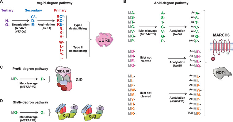
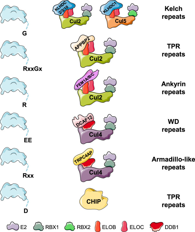
Selective protein degradation by the ubiquitin-proteasome system (UPS) is thought to be governed primarily by the recognition of specific motifs - degrons - present in substrate proteins. The ends of proteins - the N- and C-termini - have unique properties, and an important subset of protein-protein interactions involve the recognition of free termini. The first degrons to be discovered were located at the extreme N-terminus of proteins, a finding which initiated the study of the N-degron (formerly N-end rule) pathways, but only in the last few years has it emerged that a diverse set of C-degron pathways target analogous degron motifs located at the extreme C-terminus of proteins. In this minireview we summarise the N-degron and C-degron pathways currently known to operate in human cells, focussing primarily on those that have been discovered in recent years. In each case we describe the cellular machinery responsible for terminal degron recognition, and then consider some of the functional roles of terminal degron pathways. Altogether, a broad spectrum of E3 ubiquitin ligases mediate the recognition of a diverse array of terminal degron motifs; these degradative pathways have the potential to influence a wide variety of cellular functions.

Keywords: C-degron pathways; E3 ubiquitin ligases; N-degron pathways; degron; protein termini; ubiquitin proteasome system.


Conflict of interest statement

The authors declare that there are no competing interests associated with the manuscript.

Figures

Figure 1.
Figure 1.. N-degron pathways.
(A) Arg/N-degron pathway. Substrate recognition by UBR family E3 ubiquitin ligases is best understood for UBR1, which harbours two distinct substrate binding sites: one accommodates the positively charged primary type I destabilising residues (R, K and H) [71,72], whilst the second recognises the bulky hydrophobic primary type II destabilising residues (W, Y, F, L, I) [73]. Specificity for the remaining N-terminal residues comes as a result of further N-terminal processing pathways: the tertiary destabilising residues (N and Q) can be deamidated to form the secondary destabilising residues (D and E) [74,75], which are subject to N-terminal arginylation by ATE1 [76]. Oxidised cysteine (C*) is also subject to N-terminal arginylation [51]. (B) Ac/N-degron pathway. In certain contexts, acetylated N-termini can serve as degrons. It is estimated that up to 80% of all human proteins are N-terminally acetylated to some extent by N-acetyltransferase (Nat) enzymes, with the degree of acetylation varying depending on the sequence context [31]. (C) Pro/N-degron pathway. The GID E3 ligase complex targets N-terminal proline degrons. (D) Gly/N-degron pathway. Two Cul2 complexes target N-terminal glycine degrons via the substrate adaptors ZYG11B and ZER1.
Figure 2.
Figure 2.. C-degron pathways.
C-terminal degrons are targeted by a diverse array of E3 ubiquitin ligases, all of which employ tandem repeat domains to facilitate degron recognition. Elongin-B (ELOB) and Elongin-C (ELOC) bridge the interaction between the substrate adaptor and Cul2; DNA damage-binding protein 1 (DDB1) functions similarly in the assembly of Cul4 complexes. The full APPBP2 degron can be defined as Rx[2–4]Gx[0–3], with RxxGx and RxxGxx serving as optimal motifs.

References

    1. Kwon Y.T. and Ciechanover A. (2017) The ubiquitin code in the ubiquitin-proteasome system and autophagy. Trends Biochem. Sci. 42, 873–886 10.1016/j.tibs.2017.09.002 - DOI - PubMed
    1. Hershko A. and Ciechanover A. (1998) The ubiquitin system. Annu. Rev. Biochem. 67, 425–479 10.1146/annurev.biochem.67.1.425 - DOI - PubMed
    1. Pickart C.M. and Eddins M.J. (2004) Ubiquitin: structures, functions, mechanisms. Biochim. Biophys. Acta 1695, 55–72 10.1016/j.bbamcr.2004.09.019 - DOI - PubMed
    1. Dye B.T. and Schulman B.A. (2007) Structural mechanisms underlying posttranslational modification by ubiquitin-like proteins. Annu. Rev. Biophys. Biomol. Struct. 36, 131–150 10.1146/annurev.biophys.36.040306.132820 - DOI - PubMed
    1. Zheng N. and Shabek N. (2017) Ubiquitin ligases: structure, function, and regulation. Annu. Rev. Biochem. 86, 129–157 10.1146/annurev-biochem-060815-014922 - DOI - PubMed

Publication types

MeSH terms