. 2020 Oct 20;173(8):614-622.
doi: 10.7326/M20-2889. Epub 2020 Jul 6.

Clinical Validity of Serum Antibodies to SARS-CoV-2 : A Case-Control Study

Affiliations

Clinical Validity of Serum Antibodies to SARS-CoV-2 : A Case-Control Study

Giorgio Caturegli et al. Ann Intern Med. .

Abstract

Background: The clinical utility of severe acute respiratory syndrome coronavirus 2 (SARS-CoV-2) antibodies remains undefined.

Objective: To determine the clinical validity and utility of SARS-CoV-2 antibodies.

Design: Case-control study.

Setting: First month of testing for coronavirus disease 2019 (COVID-19) by using a nucleic acid amplification test (NAAT) on nasopharyngeal swabs at the Johns Hopkins Hospital, Baltimore, Maryland (11 066 persons).

Participants: Of the 11 066 tested persons, 115 (1%) were hospitalized adults investigated for COVID-19. Clinical record review was performed to classify them into a COVID-19 case group (n = 60) or a non-COVID-19 control group (n = 55). The laboratory control groups comprised 513 persons not tested by NAAT: 160 healthy laboratory employees, 101 persons positive for IgG antibodies against Epstein-Barr virus capsid antigen, 215 positive for thyroperoxidase antibody, and 37 positive for rheumatoid factor.

Measurements: Serum IgG and IgA antibodies against SARS-CoV-2 spike protein were detected by using enzyme-linked immunosorbent assay.

Results: Sensitivity and specificity of the SARS-CoV-2 IgG assay were 0.976 (95% CI, 0.928 to 0.995) and 0.988 (CI, 0.974 to 0.995), respectively, when performed 14 days or later after symptom onset, but sensitivity decreased at earlier time points. Immunoglobulin G developed rapidly and was sustained at high levels throughout follow-up (up to 58 days). Antibodies to SARS-CoV-2 predicted the odds of developing acute respiratory distress syndrome, which increased by 62% (CI, 48% to 81%; P < 0.001) for every 2-fold increase in IgG. Of 11 066 NAAT-tested patients, 457 were repeatedly NAAT-negative, and serum samples were obtained for 18 such patients (6 COVID-19 case patients and 12 non-COVID-19 control patients). Antibodies were present in 5 of 6 case patients and none of the 12 control patients (P = 0.001).

Limitations: The study was retrospective and performed at a single center; the sample was small; follow-up was limited; and selection bias may have occurred.

Conclusion: Antibodies to SARS-CoV-2 demonstrate infection when measured at least 14 days after symptom onset, are associated with clinical severity, and provide valuable diagnostic support in patients who test negative by NAAT but remain clinically suspicious for COVID-19.

Primary funding source: Clinical Immunology Laboratory, Department of Pathology, Johns Hopkins Hospital.


Conflict of interest statement

Disclosures: Authors have disclosed no conflicts of interest. Forms can be viewed at www.acponline.org/authors/icmje/ConflictOfInterestForms.do?msNum=M20-2889.

Figures

Visual Abstract.
Visual Abstract.. Clinical Validity of COVID-19 Serum Antibodies
Serum antibodies develop upon infection with SARS-CoV-2 and are currently used to stratify donors of convalescent plasma and assess the prevalence of COVID-19. This study examines the use of serum antibodies to SARS-CoV-2 spike protein in the diagnosis of COVID-19.
Figure 1.
Figure 1.. Study flow diagram.
The study included 628 participants tested for serum antibodies against spike protein. The study also included clinical record review of all 558 patients with repeated nucleic acid amplification testing of nasopharyngeal swabs (34 were tested for antibodies, whereas 524 were not). COVID-19 = coronavirus disease 2019; EBV = Epstein–Barr virus; NAAT = nucleic acid amplification test; SARS-CoV-2 = severe acute respiratory syndrome coronavirus 2.
Figure 2.
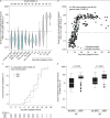
Figure 2.. Biological characteristics of serum IgG antibodies against SARS-CoV-2 spike protein in the COVID-19 case group and the laboratory control groups.
A. Violin plot showing the distribution of IgG levels, indicating median and interquartile range. The horizontal dashed line represents the manufacturer cutoff (1.1 units). B. Overall relationship between IgG levels and day post symptom onset. The white circles represent a hypogammaglobulinemic patient receiving immunosuppressive therapy due to kidney transplant. C. Kaplan–Meier survival function of IgG and IgA seroconversion. D. Serum IgG and IgA antibody levels, stratified by the presence or absence of ARDS. ARDS = acute respiratory distress syndrome; COVID-19 = coronavirus disease 2019; EBV = Epstein–Barr virus; HCW = health care worker; RF = rheumatoid factor; SARS-CoV-2 = severe acute respiratory syndrome coronavirus 2; TPO = thyroperoxidase.
Appendix Figure.
Appendix Figure.. Additional study characteristics.
Top. Distribution of follow-up time in 49 of the 60 COVID-19 patients for whom longitudinal serum samples were available. Middle. Linear relationship between serum IgG and IgA levels. Open circles indicate patients who exclusively converted IgA but not IgG before day 12 post symptom onset. Bottom. Distribution of time between first and last NAAT in 558 patients tested repeatedly. COVID-19 = coronavirus disease 2019; IQR = interquartile range; NAAT = nucleic acid amplification test; SARS-CoV-2 = severe acute respiratory syndrome coronavirus 2.
Figure 3.
Figure 3.. Integration of clinical data and molecular testing with serum antibodies to SARS-CoV-2 spike protein.
Top. Classification of 11 066 patients undergoing nasopharyngeal NAAT during the first month of COVID-19 at the Johns Hopkins Hospital, by NAAT result and number per patient. Middle. Classification of NAAT results in 558 repeatedly tested patients. Bottom. Correlation between antibody and NAAT results, classified dichotomously as ever positive or negative, in 115 hospitalized patients. Closed circles indicate COVID-19 case patients; open circles indicate non-COVID-19 hospital control patients. The top left quadrant shows the 8 patients who tested positive by NAAT and negative by antibodies (the immunosuppressed transplant patient and 7 patients with serum collection between 2 and 9 day after symptom onset). The top right and bottom left quadrants show the correlation between NAAT and antibody results, which agreed in 102 patients (46 both positive and 56 both negative). In the lower left quadrant, the closed circle indicates the antibody- and NAAT-negative patient with COVID-19 who contributed a serum sample at day 10. The lower right quadrant indicates patients with false-negative results on NAAT who tested positive for SARS-CoV-2 antibodies. COVID-19 = coronavirus disease 2019; NAAT = nucleic acid amplification test; SARS-CoV-2 = severe acute respiratory syndrome coronavirus 2.

References

    1. Dong E, Du H, Gardner L. An interactive web-based dashboard to track COVID-19 in real time [Letter]. Lancet Infect Dis. 2020;20:533-534. [PMID: 32087114] doi:10.1016/S1473-3099(20)30120-1 - PMC - PubMed
    1. Huang C, Wang Y, Li X, et al. Clinical features of patients infected with 2019 novel coronavirus in Wuhan, China. Lancet. 2020;395:497-506. [PMID: 31986264] doi:10.1016/S0140-6736(20)30183-5 - PMC - PubMed
    1. National Center for Health Statistics, Centers for Disease Control and Prevention. COVID-19 pandemic planning scenarios. 20 May 2020. Accessed at www.cdc.gov/coronavirus/2019-ncov/hcp/planning-scenarios.html on 1 June 2020.
    1. Schaller T, Hirschbühl K, Burkhardt K, et al. Postmortem examination of patients with COVID-19. JAMA. 2020. [PMID: 32437497] doi:10.1001/jama.2020.8907 - PMC - PubMed
    1. Wu F, Zhao S, Yu B, et al. A new coronavirus associated with human respiratory disease in China. Nature. 2020;579:265-269. [PMID: 32015508] doi:10.1038/s41586-020-2008-3 - PMC - PubMed

Publication types

Substances