Review
. 2020 Aug;13(8):e007952.
doi: 10.1161/CIRCEP.119.007952. Epub 2020 Jul 6.

Artificial Intelligence and Machine Learning in Arrhythmias and Cardiac Electrophysiology

Affiliations
Review

Artificial Intelligence and Machine Learning in Arrhythmias and Cardiac Electrophysiology

Albert K Feeny et al. Circ Arrhythm Electrophysiol. 2020 Aug.

Abstract

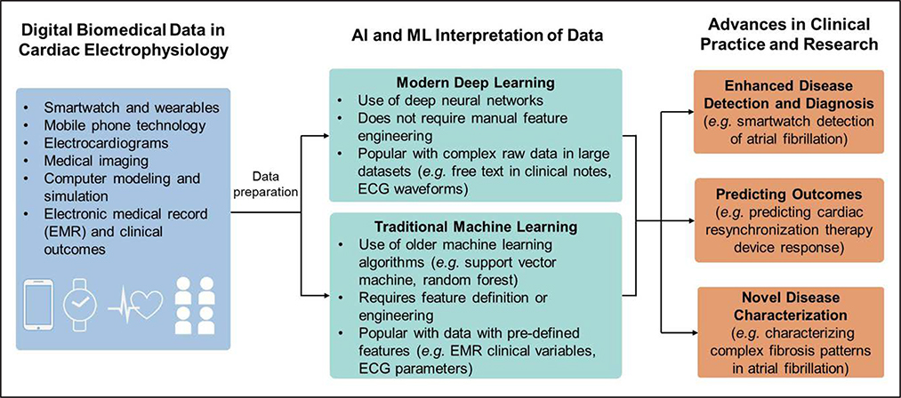
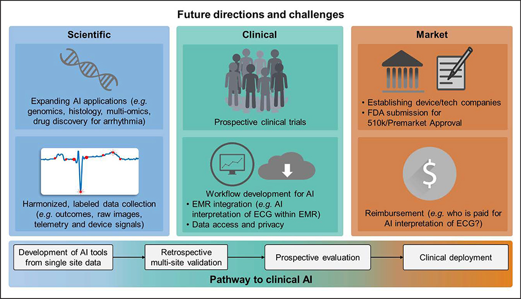
Artificial intelligence (AI) and machine learning (ML) in medicine are currently areas of intense exploration, showing potential to automate human tasks and even perform tasks beyond human capabilities. Literacy and understanding of AI/ML methods are becoming increasingly important to researchers and clinicians. The first objective of this review is to provide the novice reader with literacy of AI/ML methods and provide a foundation for how one might conduct an ML study. We provide a technical overview of some of the most commonly used terms, techniques, and challenges in AI/ML studies, with reference to recent studies in cardiac electrophysiology to illustrate key points. The second objective of this review is to use examples from recent literature to discuss how AI and ML are changing clinical practice and research in cardiac electrophysiology, with emphasis on disease detection and diagnosis, prediction of patient outcomes, and novel characterization of disease. The final objective is to highlight important considerations and challenges for appropriate validation, adoption, and deployment of AI technologies into clinical practice.

Keywords: artificial intelligence; atrial fibrillation; cardiac electrophysiology; computers; diagnosis; machine learning.


Figures

Figure 1.
Figure 1.. Overview of artificial intelligence and machine learning in cardiac electrophysiology.
A broad overview of how increasing quantities of diverse digital data in cardiac electrophysiology are being interpreted by artificial intelligence methods to generate advances in clinical practice and research. EMR indicates electronic medical record.
Figure 2.
Figure 2.. Architectures of an artificial neural network vs deep learning in ECG interpretation.
A, An example of an artificial neural network used to predict whether or not a patient will experience cardiovascular mortality, using 4 clinical features and 132 resting ECG features (intervals and amplitudes of various ECG segments). These create a 1×136 feature vector input to the neural network, represented by neurons (x1, x2, x3,…x136). The input neurons are then connected to a single fully connected hidden layer of 70 neurons (h1, h2, h3,…h70), and then ultimately connected to the output node (y), which yields a prediction score of cardiovascular mortality. The black lines between nodes represent weights, which are iteratively adjusted during the training process to minimize output prediction error. B, An example of a deep learning convolutional neural network based on the network used to predict whether or not a patient has left ventricular dysfunction from the waveforms of a 10-s 12-lead ECG. The input is the entire 12-lead ECG signal, formatted as a 12×1024 sample matrix. This network first learns temporal features within each lead, by extracting feature maps via 6 iterations of 1-dimensional convolution in the temporal axis followed by 1-dimensional pooling. Next, the network learns how the temporal features are distributed across leads by spatial feature learning via convolution across the 12 ECG leads. The resulting feature maps are flattened and passed to 2 fully connected layers (ha,1, ha,2, ha,3,…ha,64) and (hb,1, hb,2, hb,3,…hb,32), which used the learned temporal and spatial features to classify whether or not the patient has left ventricular dysfunction, as predicted in output node y.
Figure 3.
Figure 3.. Unsupervised machine learning: dimensionality reduction and k-means clustering.
Demonstration of an unsupervised machine learning approach to identify 2 subgroups of cardiac resynchronization therapy (CRT) patients based on ECG QRS complex waveforms. A, Visualization of different dimensionality reduction techniques to reduce ECG QRS waveforms from 539 CRT patients into data points in a 2-dimensional representation. Principal components analysis (PCA) is a common linear dimensionality reduction method. Other pictured techniques are nonlinear dimensionality reduction techniques: t-distributed stochastic neighbor embedding (t-SNE), and locally linear embedding (LLE). Colors of the example ECG waveforms and corresponding colors in the scatterplots indicate where each example waveform is projected in the 2-dimensional representations. B, Demonstration of k-means clustering with k=2 on the PCA representation of the ECG waveforms to create 2 clusters. The k-means algorithm is as follows: (1) k centroids are created at random locations, (2) each data point is then assigned to the nearest centroid, (3) the locations of the centroids are then updated to represent the mean location of the data points assigned to the centroid. Steps 2 and 3 are repeated over many iterations until the centroids no longer update. The centroid location and cluster assignment change over several iterations until reaching convergence.
Figure 4.
Figure 4.. Future directions for artificial intelligence (AI) in cardiac electrophysiology.
On the bottom is a typical pathway from development of AI tools to their clinical deployment. Progress thus far in electrophysiology has largely been achieved through the retrospective studies component of this pathway, with early prospective studies just beginning. There is currently significant need to evaluate and develop existing AI technologies toward clinical deployment, and potential clinical and market challenges to do so are outlined. Future scientific efforts to develop new AI tools are also outlined. EMR indicates electronic medical record; and FDA, Food and Drug Administration.

References

    1. Nygårds ME, Hulting J. An automated system for ECG monitoring. Comput Biomed Res. 1979;12:181–202. doi: 10.1016/0010-4809(79)90015-6 - DOI - PubMed
    1. Dey D, Slomka PJ, Leeson P, Comaniciu D, Shrestha S, Sengupta PP, Marwick TH. Artificial intelligence in cardiovascular imaging: JACC state-of-the-art review. J Am Coll Cardiol. 2019;73:1317–1335. doi: 10.1016/j.jacc.2018.12.054 - DOI - PMC - PubMed
    1. Martis RJ, Acharya UR, Min LC. ECG beat classification using PCA, LDA, ICA and discrete wavelet transform. Biomedical Signal Processing and Control 2013;8:437–448.
    1. Zhao Q, Zhang L. ECG Feature extraction and classification using wavelet transform and support vector machines. International Conference on Neural Networks and Brain 2005;2:1089–1092. doi: 10.1109/ICNNB.2005.1614807 - DOI
    1. Lagerholm M, Peterson C, Braccini G, Edenbrandt L, Sörnmo L. Clustering ECG complexes using hermite functions and self-organizing maps. IEEE Trans Biomed Eng. 2000;47:838–848. doi: 10.1109/10.846677 - DOI - PubMed

Publication types