. 2020 Sep 1;465(1):1-10.
doi: 10.1016/j.ydbio.2020.06.011. Epub 2020 Jul 3.

Protein phosphatase 1 regulatory subunit 35 is required for ciliogenesis, notochord morphogenesis, and cell-cycle progression during murine development

Affiliations

Protein phosphatase 1 regulatory subunit 35 is required for ciliogenesis, notochord morphogenesis, and cell-cycle progression during murine development

Danielle Archambault et al. Dev Biol. .

Abstract

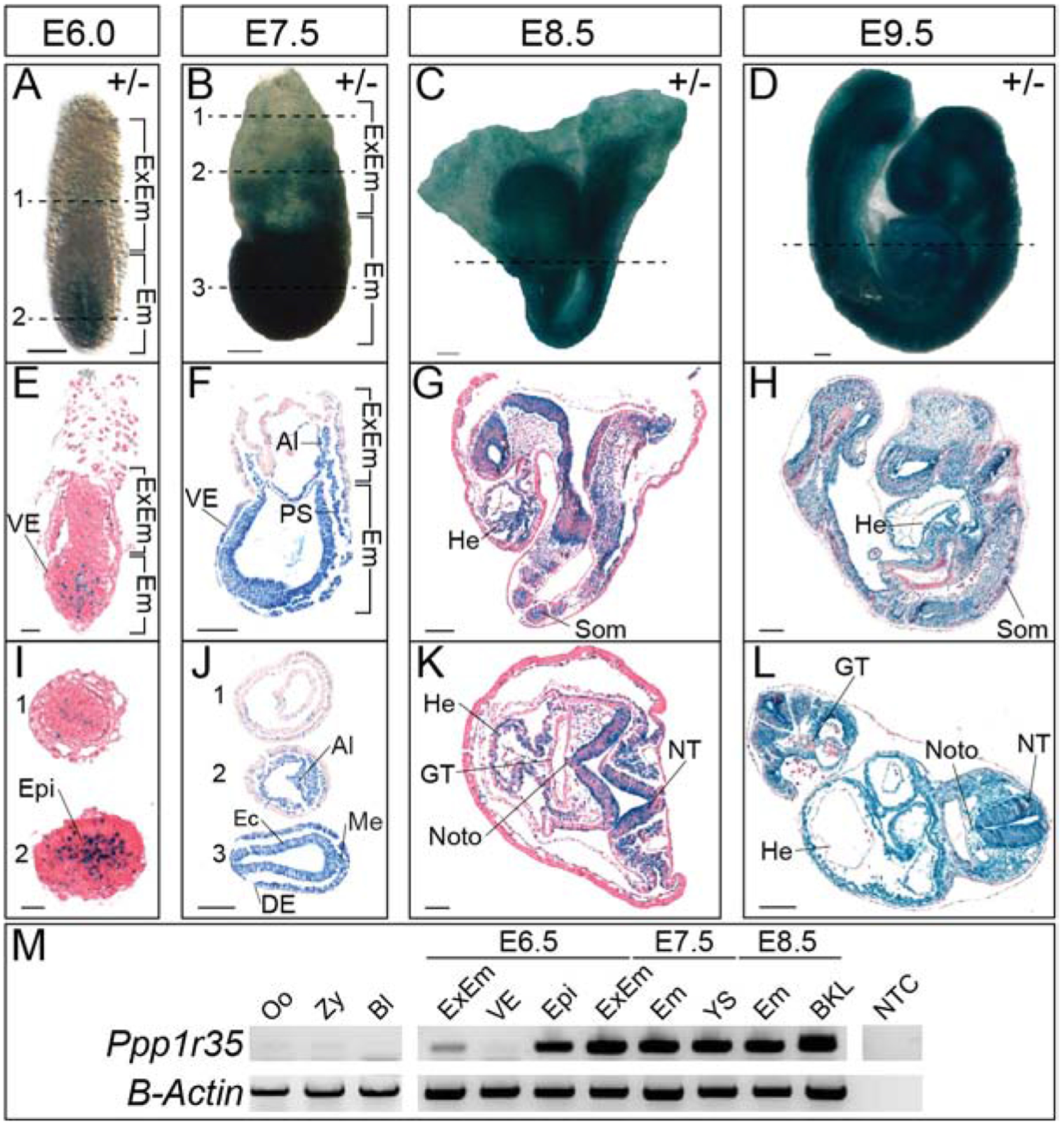
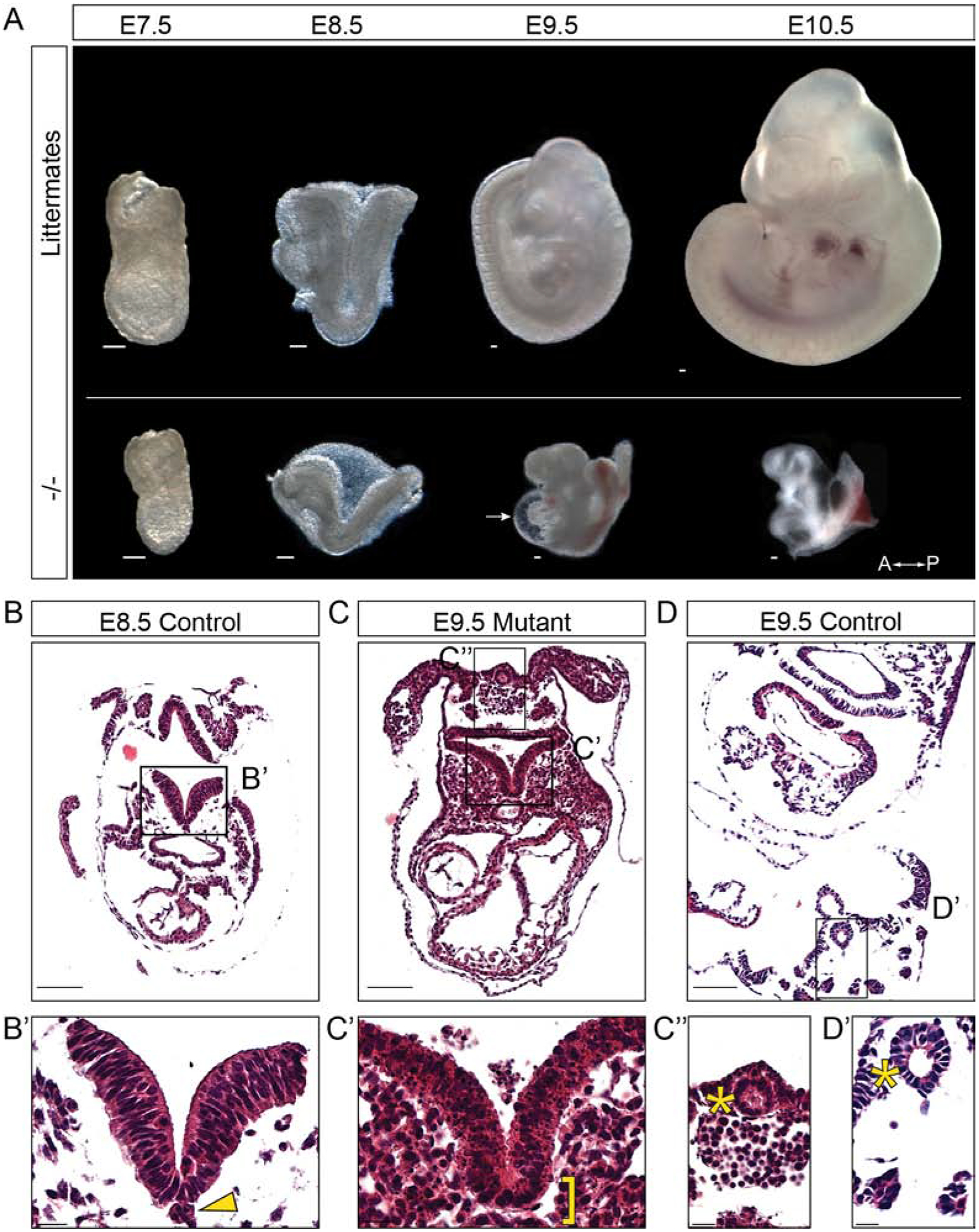
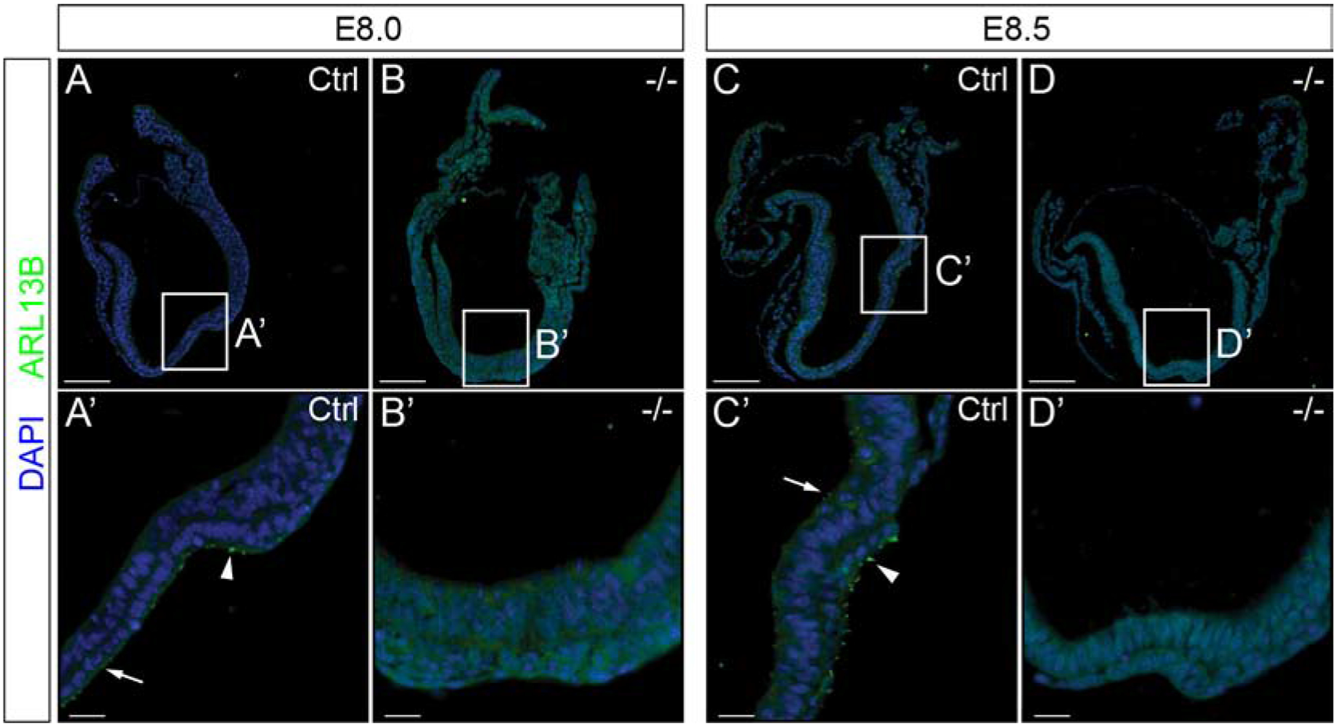
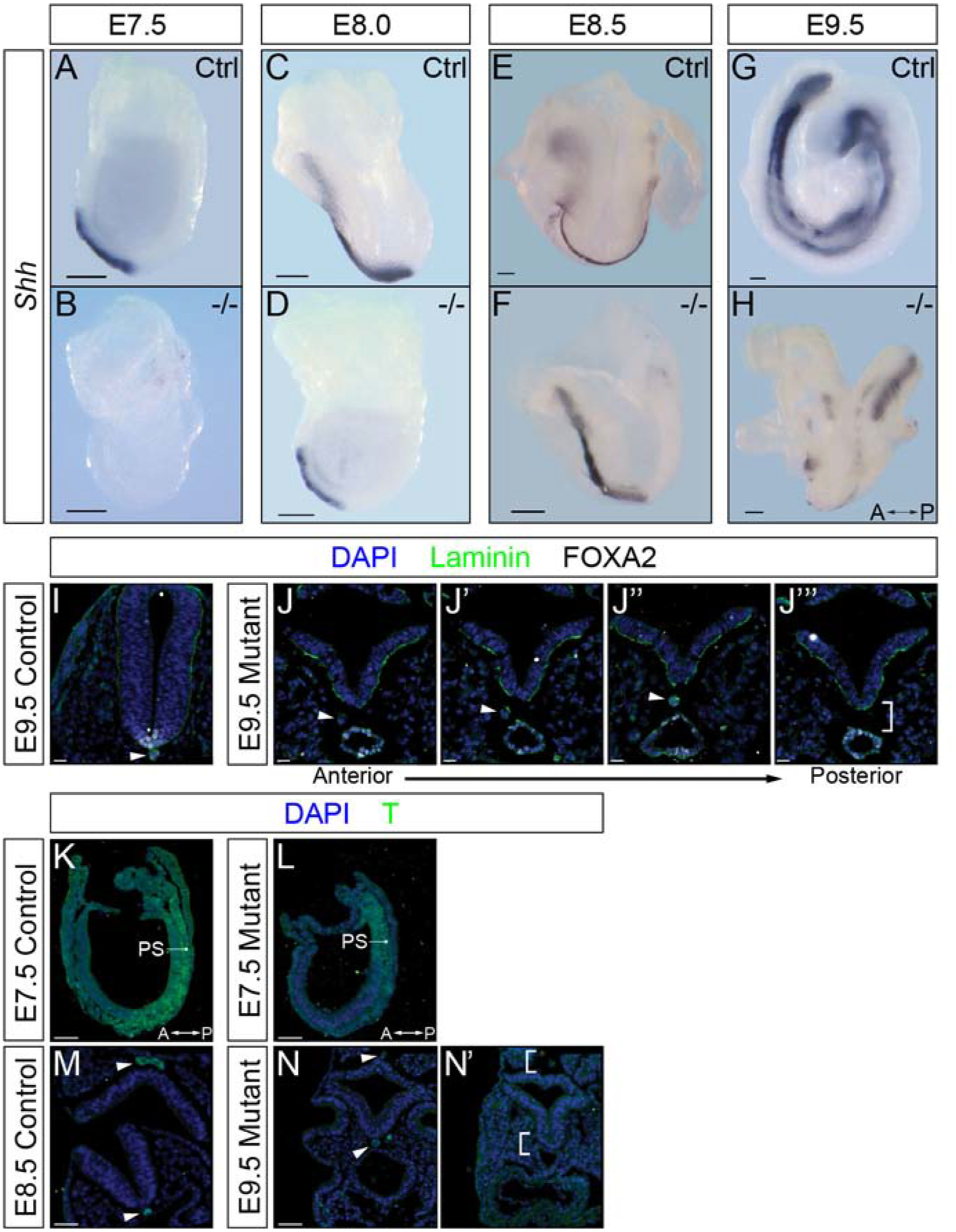
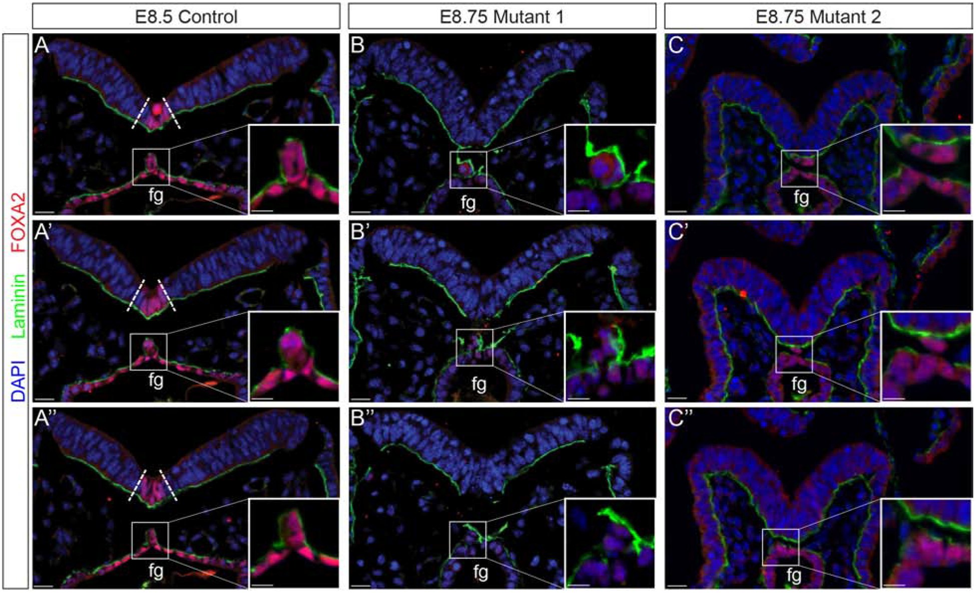
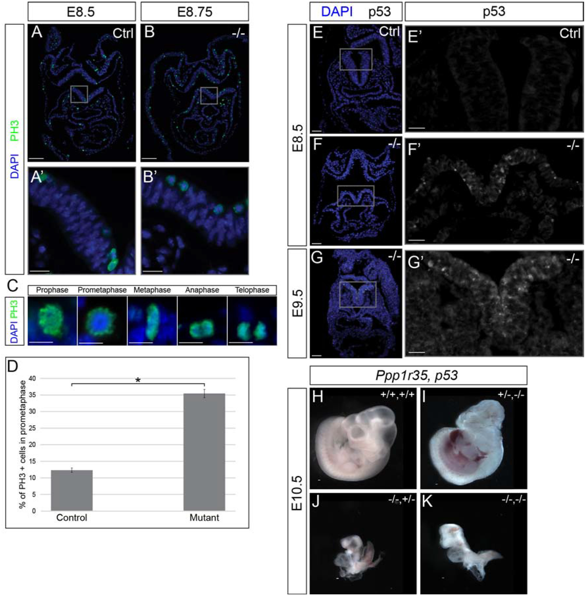
Protein phosphatases regulate a wide array of proteins through post-translational modification and are required for a plethora of intracellular events in eukaryotes. While some core components of the protein phosphatase complexes are well characterized, many subunits of these large complexes remain unstudied. Here we characterize a loss-of-function allele of the protein phosphatase 1 regulatory subunit 35 (Ppp1r35) gene. Homozygous mouse embryos lacking Ppp1r35 are developmental delayed beginning at embryonic day (E) 7.5 and have obvious morphological defects at later stages. Mutants fail to initiate turning and do not progress beyond the size or staging of normal E8.5 embryos. Consistent with recent in vitro studies linking PPP1R35 with the microcephaly protein Rotatin and with a role in centrosome formation, we show that Ppp1r35 mutant embryos lack primary cilia. Histological and molecular analysis of Ppp1r35 mutants revealed that notochord development is irregular and discontinuous and consistent with a role in primary cilia, that the floor plate of the neural tube is not specified. Similar to other mutant embryos with defects in centriole function, Ppp1r35 mutants displayed increased cell death that is prevalent in the neural tube and an increased number of proliferative cells in prometaphase. We hypothesize that loss of Ppp1r35 function abrogates centriole homeostasis, resulting in a failure to produce functional primary cilia, cell death and cell cycle delay/stalling that leads to developmental failure. Taken together, these results highlight the essential function of Ppp1r35 during early mammalian development and implicate this gene as a candidate for human microcephaly.

Keywords: Centriole; Cilia; Development; Knockout; Notochord; Ppp1r35.


Figures

Figure 1:
Figure 1:. Ppp1r35 expression during embryonic development.
X-gal stained heterozygous embryos at E6.0 (A), E7.5 (B), E8.5 (C), and E9.5 (D). Sagittal (E-H) or transverse (I-L) of X-gal stained heterozygotes at E6.0–9.5. I) At E6.0, 1 and 2 denote sections indicated by the dashed line in (A). J) At E7.5, 1–3 denote sections indicated by the dashed lines in (B). M) RT-PCR of Ppp1r35 in various tissues and at the developmental stages denoted. β-actin is a loading control. +/− denotes Ppp1r35 heterozygous embryos. ExEm; extra-embryonic, Em; embryonic, VE; visceral endoderm, Epi; epiblast, Al; allantois, PS; primitive streak, Ec; ectoderm, DE; definitive endoderm, Me; mesoderm, He; heart, Som; somites, GT; gut tube, Noto; notochord, NT; neural tube, Oo; oocyte, Zy; zygote, Bl; blastocyst, YS; yolk sac, BKL; brain-kidney-liver, NTC; no template control. Scale bars in A-D, F-H, J, and L = 100μm, E&I = 20μm, and K= 50μm.
Figure 2:
Figure 2:. Ppp1r35 knockout mouse embryos show severe morphological defects compared to their littermates.
A) Ppp1r35 mutant embryos compared to littermates collected at E7.5–10.5. Hematoxylin & eosin staining performed on transverse sections of E8.5 (B,B’) and E9.5 (D,D’) control embryos compared to an E9.5 mutant (C,C’,C”) embryo. Arrow denotes the pericardial sac. A-P indicates anterior and posterior orientation of the embryos. The arrowhead points to notochord and bracket indicates expected location of the notochord. The asterisk indicates the closed hindgut of the E9.5 mutant and E9.5 control. −/− denotes Ppp1r35 homozygous embryos. Littermates or ctrl denotes embryos wild type or heterozygous for Ppp1r35. Scale bars in A,B,C,D = 100μm and B’,C’,C”,D’ = 20μm.
Figure 3:
Figure 3:. Ppp1r35 is necessary for primary cilia formation in the developing embryo.
Embryos collected at E8.0 and E8.5 were labeled using ARL13B (green), and nuclei counterstained with DAPI (blue). Ppp1r35 E8.0 and 8.5 control embryos have ARL13B-positive cilia as expected (A,A’) and (C,C’). However, mutant embryos have a complete loss of ARL13B-positive primary cilia (B,B’) (D,D’). Arrowhead denotes nodal cilia and an arrow points to positive puncta. White box indicates the location of the node and corresponds to A’,B’,C’, and D’ respectively. −/− denotes Ppp1r35 homozygous embryos. Ctrl denotes embryos wild type or heterozygous for Ppp1r35. ARL13B mutant embryos: n=5. Scale bars for A-D = 100 μm and A’-D’ = 20 μm.
Figure 4:
Figure 4:. Shh expression and notochord are disrupted in Ppp1r35 mutant embryos at E9.5.
Whole-mount in situ hybridization to detect sonic hedgehog (Shh) expression in littermates and Ppp1r35 mutant embryos collected at E7.5 (A,B), E8.0 (C,D), E8.5 (E,F) and E9.5 (G,H). Shh mutant embryos: E7.5 n=3, E8.0 n=2, E8.5 n=3, E9.5 n=3. Immunofluorescence of transversely sectioned E9.5 embryos using laminin (green), FOXA2 (white) and nuclei counterstained with DAPI (blue) (I,J-J”‘). FOXA2 mutant embryos: n=3. T (green) and DAPI (blue) staining of sectioned E7.5 sagittal control (K), E7.5 sagittal mutant (L), E8.5 transverse control (M) and E9.5 transverse mutant (N,N’). T mutant embryos: E7.5 n=3, E9.5 n=3. A-P indicates the anterior-posterior axis of the E9.5 mutant embryo. Arrowhead indicates FOXA2 and T positive notochord. Brackets indicate expected location of the notochord. −/− denotes Ppp1r35 homozygous embryos. Ctrl denotes wild type or heterozygous Ppp1r35 embryos. Scale bars for A-H = 100 μm, I-J” = 20 μm, K-N’ = 50 μm. The arrows indicate the position of the anterior and posterior (A-P) axis.
Figure 5:
Figure 5:. Notochord morphogenesis is disrupted in Ppp1r35 mutant embryos.
Immunofluorescent analysis of laminin (green), FOXA2 (red), and nuclei counterstained with DAPI (blue). Consecutive sections through a stage-matched E8.5 control reveals a FOXA2-positive floor plate (boundary defined by dotted lines) and a contiguous notochord that has laminin distributed laterally and dorsally (A-A”). Although the two E8.75 two mutants vary slightly they both lack a FOXA2-positive floor plate and display altered notochord resolution (B-B”, C-C”). Fg; foregut. Ctrl denotes embryos wild type or heterozygous for Ppp1r35. −/− denotes Ppp1r35 homozygous embryos. Boxes indicate a higher magnification of the notochord. FOXA2-labeled mutant embryos: n=3. Scale bars = 20 μm.
Figure 6:
Figure 6:. Ppp1r35 mutants display increased apoptosis in the neural tube and a significant, embryo-wide, increase in prometaphase cells.
Immunofluorescent analysis of cell proliferation using PH3 (green), counterstained with DAPI (blue). E8.5 stage-matched control used for the E8.75 mutant embryo (A,B). PH3 mutant embryos: n=3. PH3 positive cells undergoing the stages of mitosis (C). Significantly more PH3-positive cells in prometaphase are observed in mutant embryos (compare A’ to B’, quantified in D). Analysis of active p53 (white; E-G’) and p53 with nuclei (DAPI; blue) (E,F,G). No/rare p53 positive cells are found in control E8.5 embryos (E,E’). Many p53-positive cells are observed in the neural tube of the E8.5 (F,F’) and E9.5 (G,G’) Ppp1r35 mutants. p53 mutant embryos: n=3. No obvious phenotypic rescue was observed in Ppp1r35;p53 double-knockout embryos collected at E10.5 (H-K). −/− denotes Ppp1r35 homozygous embryos. Littermates or ctrl denotes embryos wild type or heterozygous for Ppp1r35. +/+,+/+ (WT Ppp1r35,WT p53); +/−,−/− (Het Ppp1r35, Mut p53); −/−,+/− (Mut Ppp1r35, Het p53); −/−,−/− (Mut Ppp1r35, Mut p53). Scale bars for A and B = 100 μm, A’ and B’ = 20 μm, C = 10 μm, E,F,G = 50 μm, E’,F’,G’ = 20 μm, H-K = 100μm. Error bars indicate SEM. Statistically significant comparisons are indicated with a bracket and star where *P<0.001. (P= 9.31E-16).

References

    1. Pertea M, et al., Thousands of large-scale RNA sequencing experiments yield a comprehensive new human gene list and reveal extensive transcriptional noise. bioRxiv, 2018: p. 332825. - PMC - PubMed
    1. UCDavis. KOMP Repository Knockout Mouse Project. 2019; Available from: http://www.KOMP.org.
    1. Consortium IMP Gene: Ppp1r35. 2019; Available from: https://www.mousephenotype.org/data/genes/MGI:1922853.
    1. Dickinson ME, et al., High-throughput discovery of novel developmental phenotypes. Nature, 2016. 537: p. 508. - PMC - PubMed
    1. Jurand A, Some aspects of the development of the notochord in mouse embryos. J Embryol Exp Morphol, 1974. 32(1): p. 1–33. - PubMed

Publication types

Substances