Review
. 2020 Jul 24;23(7):101301.
doi: 10.1016/j.isci.2020.101301. Epub 2020 Jun 20.

Cannabinoids and Cannabinoid Receptors: The Story so Far

Affiliations
Review

Cannabinoids and Cannabinoid Receptors: The Story so Far

Fred Shahbazi et al. iScience. .

Abstract

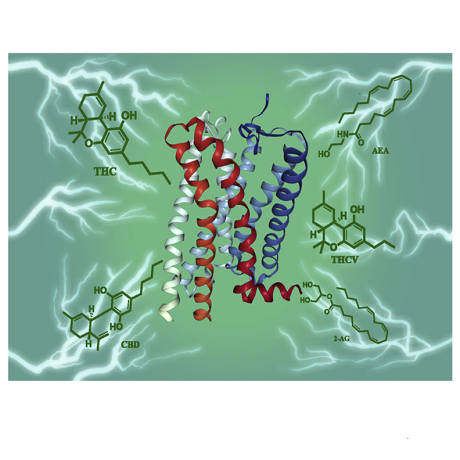
Like most modern molecular biology and natural product chemistry, understanding cannabinoid pharmacology centers around molecular interactions, in this case, between the cannabinoids and their putative targets, the G-protein coupled receptors (GPCRs) cannabinoid receptor 1 (CB1) and cannabinoid receptor 2 (CB2). Understanding the complex structure and interplay between the partners in this molecular dance is required to understand the mechanism of action of synthetic, endogenous, and phytochemical cannabinoids. This review, with 91 references, surveys our understanding of the structural biology of the cannabinoids and their target receptors including both a critical comparison of the extant crystal structures and the computationally derived homology models, as well as an in-depth discussion about the binding modes of the major cannabinoids. The aim is to assist in situating structural biochemists, synthetic chemists, and molecular biologists who are new to the field of cannabis research.

Keywords: Medical Substance; Molecular Biology; Structural Biology; Supramolecular Chemistry.


Figures

None
Graphical abstract
Figure 1
Figure 1
Structure of Cannabinoids Archetypal of Their Structural Families
Figure 2
Figure 2
Schematic Representation of Signal Transduction by Ligand Interactions with the GPCRs The state of the receptor in each panel is described therein.
Figure 3
Figure 3
Structure of Synthetic Analogs AM6538, TNB, AM11542, FUB, AM10257, AM12033, and WIN55,212-2
Figure 4
Figure 4
Comparison of CB1 Binding to Different Ligands Analysis of the ligand binding pocket (A) AM6538@CB1 (5TGZ) and (B) AM11542@CB1 (5XRA). (C) Comparison of the agonist-bound (blue) and antagonist-bound (light blue) CB1. (D) Superposition of FUB@CB1 (navy) and AM11542@CB1 (blue). Data used to prepare the figures were obtained from Hua et al. (2016), Figures 4A; Hua et al. (2017), Figure 4B; and Krishna Kumar et al. (2019), Figure 4D.
Figure 5
Figure 5
Comparison of the Ligand-Binding Modes of CB1 and CB2 (A and B) (A) Superposition of AM6538@CB1 (5TGZ) (blue) and AM10257@CB2 (5ZTY) (orange) ligand-binding pockets and (B) conformational difference of F200 and W258 in antagonist bound CB2 and CB1. (C) Comparison of the “toggle switch” residue conformation in agonist AM12033@CB2 (6KPC) (gold) and AM10257@CB2 (5ZTY) (orange). (D–G) (D) WIN55212-2@CB2 (6PT0) (yellow) orthosteric pocket shows the direct contact of the ligand with residues F1173.36 and W2586.48 compared with AM10257@CB2 (orange). Comparison of the gross morphology of the receptors: (E) active CB1 and CB2 (F), active CB1 and inactive CB2, and (G) inactive CB1 and CB2. Data used to prepare the figures were obtained from Li et al. (2019), Figures 5A and 5B; Hua et al. (2020), Figures 5C and 5E–5G; and Xing et al. (2020), Figure 5D.
Figure 6
Figure 6
General CB1 Receptor Inverse Agonist Pharmacophore Model in Relation to the Putative CB1 Receptor Side Chain Residues in Receptor-Ligand Interactions Figure adapted from Lange and Kruse (2005).
Figure 7
Figure 7
THC and AEA Bind to Both CB1 and CB2 Nonselective agonists THC and AEA bound to (A) CB1 and (B) CB2 in complexes characterized by the lowest energies; both top-down and side-on views are provided. Interactions involving π orbitals are shown as orange solid lines, and hydrogen bonds are shown as green dashed lines. Figure reproduced from Latek et al., (2011), with permission.
Figure 8
Figure 8
THC and TNB Adopt Different Comformations when Bound to CB1 (A–C) (A) Chemical structures and predicted binding poses of THC with inactive structure, (A) PBD: 5TGZ and (B) PDB: 5U09 (15) (transparent magenta sticks), and (C) with active structure PDB: 5XRA. THC (pink) and TNB (light green). Data used to prepare the figures were obtained from Hua et al., 2016 Figure 8A; Shao et al. (2016), Figure 8B; and Hua et al., 2017, Figure 8C.
Figure 9
Figure 9
CBD and THC May Bind Simultaneously to CB1 (A–C) (A) Orthosteric site (O) and the three potential allosteric binding pockets in CB1 PDB: 5TGZ. (B) Solvent rearrangement around CBD in the agonist-bound CB1. CBD (orange), THC (yellow), and water molecules within ≥3 Å in the active conformation of the CB1R at 0, 25, and 50 ns of simulation; (C) coordinated movement observed in the active conformation of CB1 during 50 ns of simulation. Data used to prepare (A) was obtained from Sabatucci et al., 2018; Figures 9B and 9C are reproduced from Chung et al. (2019) under Creative Commons Rights.
Figure 10
Figure 10
Binding Poses of CB1 Ligands after 1-μs Molecular Dynamics Simulations (A–G) THC and THCV were bound to the (A) inactive and (B) active conformations of CB1 receptor. Docking pose of (C) THC, (D) THCV, (E) THCB, and (F and G) THCP in complex with CB1 receptor (PDB ID: 5XRA). Data used to prepare Figures 10A and 10B were obtained from Jung et al. (2018), and for Figures 10C–10G from Citti et al. (2019a, .
Figure 11
Figure 11
Endogenous Cannabinoids Interact with the CB2 Receptor (A) Molecular dynamics simulations of 2-AG binding to the membrane-embedded CB2 receptor. Predicted binding poses of (B) AEA and (C) 2-AG. Figure 11A is reproduced from Hurst et al. (2010) with permission; data used to generate Figures 11B and 11C were obtained from Hua et al.

References

    1. Baker D., Pryce G., Giovannoni G., Thompson A.J. The therapeutic potential of cannabis. Lancet Neurol. 2003;2:291–298. - PubMed
    1. Bouaboula M., Poinot-Chazel C., Marchand J., Canat X., Bourrié B., Rinaldi-Carmona M., Calandra B., Le Fur G., Casellas P. Signaling pathway associated with stimulation of CB2 peripheral cannabinoid receptor. Eur. J. Biochem. 1996;237:704–711. - PubMed
    1. Bow E.W., Rimoldi J.M. The structure–function relationships of classical cannabinoids: CB1/CB2 modulation. Perspect. Medicin. Chem. 2016;8:17–39. - PMC - PubMed
    1. Cherezov V., Rosenbaum D.M., Hanson M.A., Rasmussen S.G.F., Thian F.S., Kobilka T.S., Choi H.-J., Kuhn P., Weis W.I., Kobilka B.K. High-Resolution crystal structure of an engineered human β2-adrenergic G protein–coupled receptor. Science. 2007;318:1258–1265. - PMC - PubMed
    1. Chien E.Y.T., Liu W., Zhao Q., Katritch V., Won Han G., Hanson M.A., Shi L., Newman A.H., Javitch J.A., Cherezov V. Structure of the human dopamine D3 receptor in complex with a D2/D3 selective antagonist. Science. 2010;330:1091–1095. - PMC - PubMed