

A Simple Method for the Production of Human Skin Equivalent in 3D, Multi-Cell Culture
- PMID: 32629914
- PMCID: PMC7369873
- DOI: 10.3390/ijms21134644
A Simple Method for the Production of Human Skin Equivalent in 3D, Multi-Cell Culture
Abstract
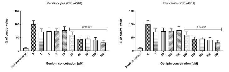
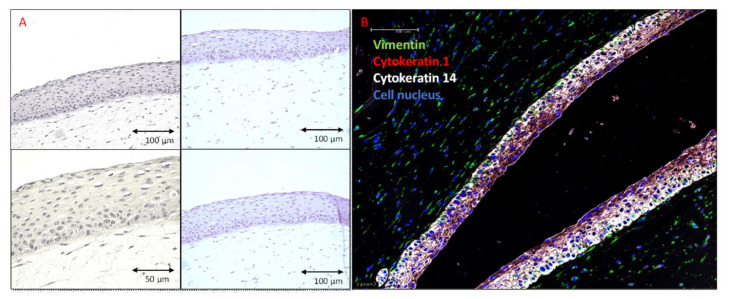
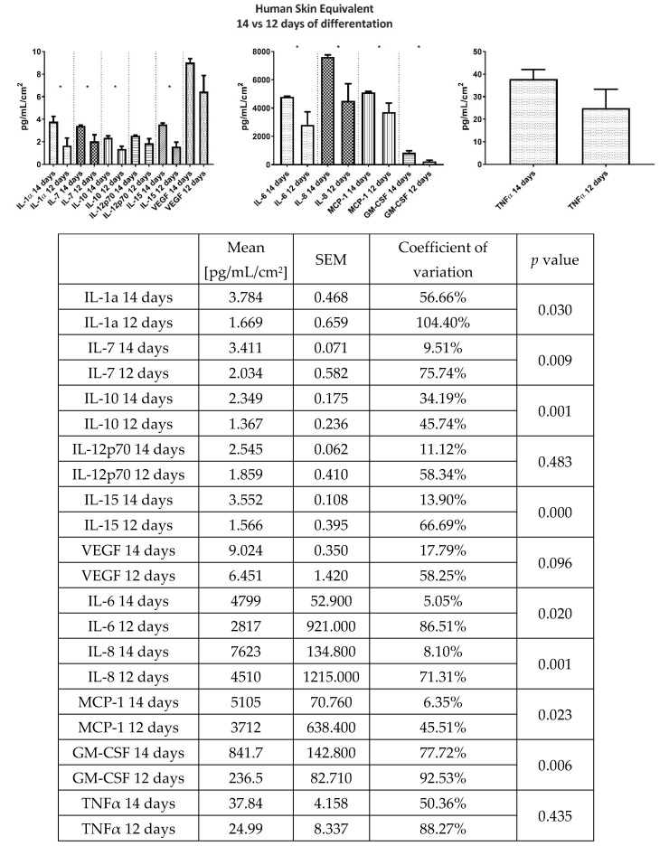
An important problem for researchers working in the field of dermatology is the preparation of the human skin equivalent (HSE). Here, we describe a simple and reliable protocol for preparing a skin model from the commercially available cell lines: keratinocytes, fibroblasts, and melanocytes. Importantly, in our 3D model, the keratinocytes are diverse that brings this model closer to the natural skin. For the production of HSE, we used available primary PCS-200-010, PCS-201-010, PCS-200-013, and immortalized CRL-4048 and CRL-4001 cell lines. We used genipin, which is necessary for collagen cross-linking and studied its cytotoxicity for keratinocytes and fibroblasts. The addition of 20 μM genipin reduced the shrinkage of the collagen in the constructs from 59% to 24% on day 12 of the culture of the construct. A higher concentration (80-200 µM) of genipin reduced shrinkage by 14% on average. Genipin in concentration 10 μM and below was not cytotoxic to the keratinocytes, and 150 μM and below to the fibroblasts. Hematoxylin and eosin staining showed that the morphology of HSEs was identical to that of native human skin. The immunohistochemical staining of the constructs showed the presence of vimentin-positive fibroblasts in the skin layer, while the melanocytes were in the epidermis and in the basal layer. We observed that the longer differentiation of constructs led to the higher secretion of GM-CSF, IL-10, IL-15, IL-1α, IL-6, IL-7, IL-8, and MCP-1. We also observed that the longer time of differentiation led to a more stable secretion of all analytes, which was reflected in the coefficient of variation. We described here a simple, reliable, and cost-effective production of the full-thickness human skin equivalents that can be used in the research and industry. With the global trend to decrease animal use for the research and testing, our HSE could be a useful testing tool and an alternative research model.
Keywords: 3D model; commercially available cell lines; cytokines; differentiated keratinocytes; skin equivalent.
Conflict of interest statement
The authors certify that there is no conflict of interest with any financial organization regarding the material discussed in the manuscript.
Figures
References
-
- Duval C., Chagnoleau C., Pouradier F., Sextius P., Condom E., Bernerd F. Human Skin Model Containing Melanocytes: Essential Role of Keratinocyte Growth Factor for Constitutive Pigmentation—Functional Response to α-Melanocyte Stimulating Hormone and Forskolin. Tissue Eng. Part C Methods. 2012;18:947–957. doi: 10.1089/ten.tec.2011.0676. - DOI - PubMed
-
- Hachiya A., Sriwiriyanont P., Kaiho E., Kitahara T., Takema Y., Tsuboi R. An In Vivo Mouse Model of Human Skin Substitute Containing Spontaneously Sorted Melanocytes Demonstrates Physiological Changes after UVB Irradiation. J. Investig. Dermatol. 2005;125:364–372. doi: 10.1111/j.0022-202X.2005.23832.x. - DOI - PubMed
MeSH terms
Substances
Grants and funding
LinkOut - more resources
Full Text Sources
Miscellaneous
