Review
. 2020 Jun 30;21(13):4682.
doi: 10.3390/ijms21134682.

Wnt/β-Catenin Signaling in Oral Carcinogenesis

Affiliations
Review

Wnt/β-Catenin Signaling in Oral Carcinogenesis

Montserrat Reyes et al. Int J Mol Sci. .

Abstract

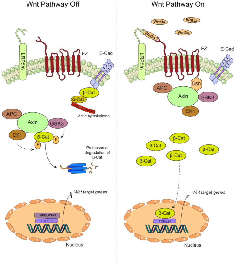
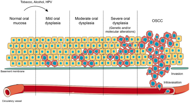
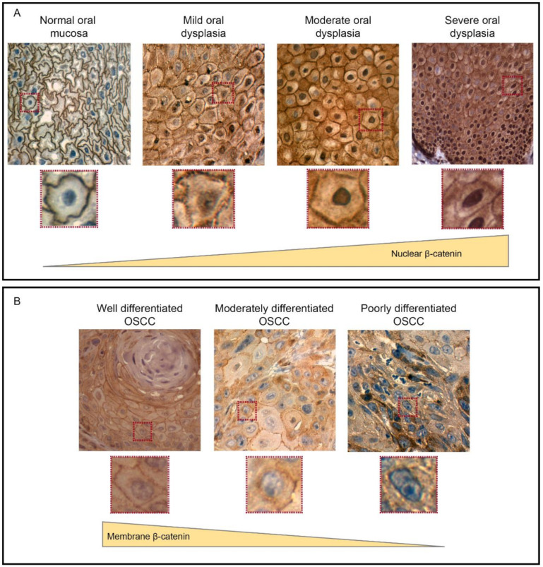
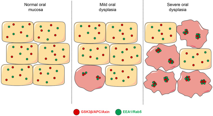
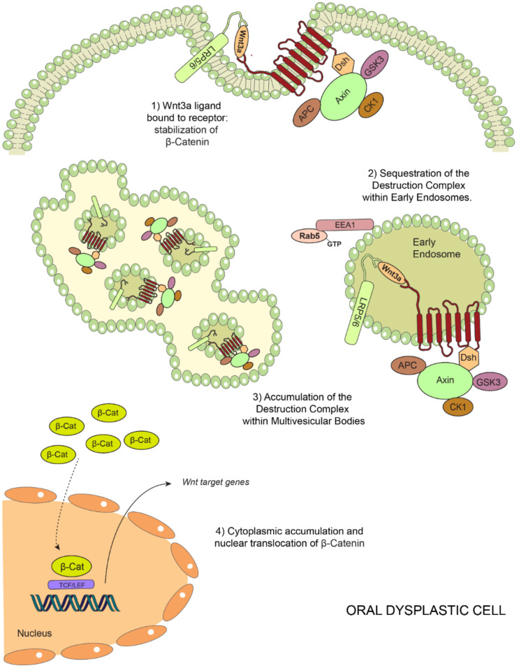
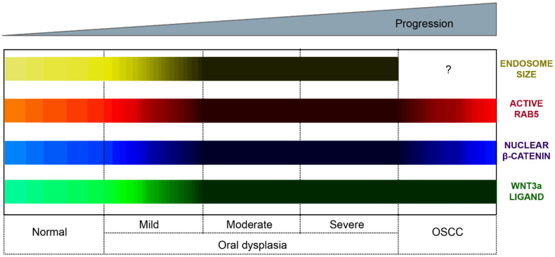
Oral carcinogenesis is a complex and multifactorial process that involves cumulative genetic and molecular alterations, leading to uncontrolled cell proliferation, impaired DNA repair and defective cell death. At the early stages, the onset of potentially malignant lesions in the oral mucosa, or oral dysplasia, is associated with higher rates of malignant progression towards carcinoma in situ and invasive carcinoma. Efforts have been made to get insights about signaling pathways that are deregulated in oral dysplasia, as these could be translated into novel markers and might represent promising therapeutic targets. In this context, recent evidence underscored the relevance of the Wnt/β-catenin signaling pathway in oral dysplasia, as this pathway is progressively "switched on" through the different grades of dysplasia (mild, moderate and severe dysplasia), with the consequent nuclear translocation of β-catenin and expression of target genes associated with the maintenance of representative traits of oral dysplasia, namely cell proliferation and viability. Intriguingly, recent studies provide an unanticipated connection between active β-catenin signaling and deregulated endosome trafficking in oral dysplasia, highlighting the relevance of endocytic components in oral carcinogenesis. This review summarizes evidence about the role of the Wnt/β-catenin signaling pathway and the underlying mechanisms that account for its aberrant activation in oral carcinogenesis.

Keywords: Rab5; destruction complex; dysplasia; endosome; oral cancer; β-catenin.


Conflict of interest statement

The authors declare no conflicts of interest. The funders had no role in the design of the study; in the collection, analyses or interpretation of data; in the writing of the manuscript or in the decision to publish the results.

Figures

Figure 1
Figure 1
The canonical Wnt pathway. (Left) In the absence of extracellular Wnt ligands, the transmembrane receptors Frizzled (FZ) and the coreceptors LDL (low-density lipoprotein) receptor related protein 5/6 (LRP 5/6) are unable to associate at the plasma membrane, yielding an “off” state of the pathway. During this off state, β-catenin (β-Cat) is mainly found at cell-cell adhesion complexes, bridging together the intercellular adhesion molecule E-Cadherin (E-cad) and the actin cytoskeleton via interaction with α-catenin (α-Cat). In the cytoplasm, β-catenin is rapidly targeted for proteasomal degradation by the so-called “destruction complex”, which is composed by adenomatous polyposis coli (APC), axin, casein kinase 1 (CK1) and glycogen synthase kinase 3β (GSK3β). The targeting of β-catenin for degradation is based on sequential phosphorylation by CK1 and GSK3β. In these conditions, β-catenin cannot translocate to the nucleus, and the transcription of the target genes is repressed by GROUCHO, which is bound to the TCF/LEF promoters. (Right) Secreted Wnt ligands, such as Wnt3a, are recognized by both FZ and LRP5/6, switching “on” the pathway. The destruction complex is then recruited to the plasma membrane via interaction with the FZ receptor, allowing the cytoplasmic accumulation of β-catenin, which is now available for translocation to the nucleus, where it binds the TCF/LEF promoter by displacing GROUCHO, allowing the transcription of Wnt target genes. Particularly, in oral carcinogenesis, this pathway is “switched on” by the increased secretion of Wnt3a, stabilization of β-catenin and the expression of target genes such as cyclin D1 and survivin (see main text for details).
Figure 2
Figure 2
Oral carcinogenesis. Normal oral mucosa is a stratified layer of epithelial cells arranged over a basement membrane that separates epithelial cells from connective tissue and blood vessels. When oral mucosa is challenged with external stressors, such as tobacco, alcohol or human papilloma virus (HPV) infection, cells in the deepest layers undergo morphological alterations in shape and size. This novel state represents an adaptation response against a harmful stimulation, which is known as oral dysplasia. Oral dysplasia might be categorized as mild, moderate or severe, according with the extension of the lesion and the presence of molecular markers induced as result of the altered genetic expression. Oral dysplasia is considered the previous stage before oral squamous cell carcinoma (OSCC) and the strongest predictor of malignant transformation to cancer. During OSCC, massive phenotypic changes affect all epithelial layers, and it is extended over the tissue border, with ruptures of the basement membrane, in a process that allows the invasion of the connective tissue and incorporation into blood vessels (intravasation).
Figure 3
Figure 3
The expression and localization of β-catenin during oral carcinogenesis. (A) Immunohistochemistry of β-catenin in human oral samples, which revealed that β-catenin is mainly found at the plasma membrane of epithelial cells in normal oral mucosa samples. However, in oral dysplasia, β-catenin is mostly accumulated at the cytoplasm and nucleus of epithelial cells. Importantly, the nuclear detection of β-catenin is progressively increased according with the degree of dysplasia, with the strongest detection in severe and moderate oral dysplasia (zoomed images are shown in lower panels). (B) In OSCC, nuclear β-catenin levels are lower than oral dysplasia. On the other hand, plasma membrane-associated β-catenin decreases when shifting from well-differentiated to moderate and poorly differentiated OSCC (zoomed images are shown in the lower panels). This figure was modified with permission from “Increased nuclear β-catenin expression in oral potentially malignant lesions: A marker of epithelial dysplasia”, Reyes M. et al. 2015, Med Oral Patol Oral Cir Bucal.
Figure 4
Figure 4
Early endosomes and their colocalization with components of the β-catenin destruction complex. This figure depicts a model that summarizes different aspects related to the mechanisms involved in Wnt/β-catenin signaling in oral dysplasia. First, early endosomes (shown in green, EEA1- and Rab5-positive) are progressively enlarged throughout the different stages of oral dysplasia in a manner that their co-localization with the components of the β-catenin destruction complex is also augmented during the progression of oral dysplasia. Specifically, the proteins APC, axin and GSK3β have been shown to increasingly colocalize with EEA1- and Rab5-positive early endosomes.
Figure 5
Figure 5
Model of endosomal sequestration of the destruction complex in oral dysplasia. Binding of Wnt3a to Frizzled (FZ) and LRP5/6 leads to the recruitment of the destruction complex to the FZ-LRP5/6 receptor complex at the plasma membrane. This is followed by a decreased proteasomal degradation of β-catenin (1). Ligand binding and subsequent posttranslational modifications (not described in this scheme) lead to endocytosis of this supramolecular complex, also known as the “Wnt signalosome complex”, and subsequent trafficking en route to early endosomes and multivesicular bodies in a Rab5-dependent manner (2). In oral dysplasia, high Wnt3a levels and increased Rab5 activity lead to the enhanced sequestration of the destruction complex within EEA1-positive early endosomes. Consequently, higher levels of components of the destruction complex are detected in multivesicular bodies (3). These events ultimately lead to a more robust stabilization of β-catenin in the cytoplasm and a consequent nuclear translocation in order to bind TCF/LEF factors, activating the transcription of the target genes (4).
Figure 6
Figure 6
Subcellular and molecular events in oral carcinogenesis. This scheme provides a summary of subcellular and molecular changes observed during oral carcinogenesis. These changes include the Wnt3a expression, nuclear detection of β-catenin, endosome enlargement and the activation status of Rab5 GTPase. We propose a model whereby oral carcinogenesis is associated with the progressive expression of Wnt3a and the consequent stabilization and nuclear translocation of β-catenin. These events are accompanied by the continuous activation of Rab5-GTPase, endosome enlargement and the increased sequestration of the destruction complex within endosomes (see main text for details). Color codes represent the Wnt3a expression (green bar), nuclear β-catenin detection (blue bar), Rab5 activity (red bar) and early endosome size (yellow bar). The lighter the color, the lower the expression/detection/activity/size. The darker the color, the higher the expression/detection/activity/size for each case. The Wnt3a representation summarizes evidence obtained from immunohistochemical analyses in clinical samples and in vitro measurements of ligand secretions in cell culture models. The nuclear β-catenin representation summarizes evidence obtained in both clinical samples and cell culture models. The nuclear detection progressively increases from normal oral mucosa through the different stages of oral dysplasia (mild, moderate and severe); however, the nuclear detection of this protein decreases in OSCC. The Rab5 activity (Rab5-GTP levels) has been measured in cell culture models and is shown substantially increased in dysplastic oral keratinocytes (models of moderate/severe dysplasia) in comparison with nondysplastic oral keratinocytes and OSCC cells. The endosomal size (early endosomes) has been measured by tissue immunofluorescence in clinical samples of normal mucosa and mild, moderate and severe dysplasia, as well as in cell culture models, using different markers of early endosomes (the interrogation symbol indicates that this has not been explored in OSCC).

References

    1. Haddad R.I., Shin D.M. Recent advances in head and neck cancer. N. Engl. J. Med. 2008;359:1143–1154. doi: 10.1056/NEJMra0707975. - DOI - PubMed
    1. Leemans C.R., Braakhuis B.J., Brakenhoff R.H. The molecular biology of head and neck cancer. Nat. Rev. Cancer. 2011;11:9–22. doi: 10.1038/nrc2982. - DOI - PubMed
    1. Neville B.W., Day T.A. Oral cancer and precancerous lesions. CA. Cancer J. Clin. 2002;52:195–215. doi: 10.3322/canjclin.52.4.195. - DOI - PubMed
    1. Polanska H., Raudenska M., Gumulec J., Sztalmachova M., Adam V., Kizek R., Masarik M. Clinical significance of head and neck squamous cell cancer biomarkers. Oral Oncol. 2014;50:168–177. doi: 10.1016/j.oraloncology.2013.12.008. - DOI - PubMed
    1. Genden E.M., Ferlito A., Silver C.E., Takes R.P., Suarez C., Owen R.P., Haigentz M., Stoeckli S.J., Shaha A.R., Rapidis A.D., et al. Contemporary management of cancer of the oral cavity. Eur. Arch. Otorhinolaryngol. 2010;267:1001–1017. doi: 10.1007/s00405-010-1206-2. - DOI - PMC - PubMed