Review
. 2020 Jun 30;21(13):4688.
doi: 10.3390/ijms21134688.

Vascular Homeostasis and Inflammation in Health and Disease-Lessons from Single Cell Technologies

Affiliations
Review

Vascular Homeostasis and Inflammation in Health and Disease-Lessons from Single Cell Technologies

Olga Bondareva et al. Int J Mol Sci. .

Abstract

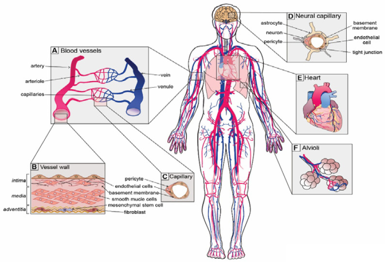
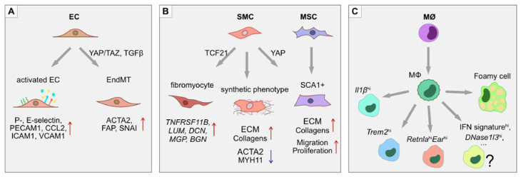
The vascular system is critical infrastructure that transports oxygen and nutrients around the body, and dynamically adapts its function to an array of environmental changes. To fulfil the demands of diverse organs, each with unique functions and requirements, the vascular system displays vast regional heterogeneity as well as specialized cell types. Our understanding of the heterogeneity of vascular cells and the molecular mechanisms that regulate their function is beginning to benefit greatly from the rapid development of single cell technologies. Recent studies have started to analyze and map vascular beds in a range of organs in healthy and diseased states at single cell resolution. The current review focuses on recent biological insights on the vascular system garnered from single cell analyses. We cover the themes of vascular heterogeneity, phenotypic plasticity of vascular cells in pathologies such as atherosclerosis and cardiovascular disease, as well as the contribution of defective microvasculature to the development of neurodegenerative disorders such as Alzheimer's disease. Further adaptation of single cell technologies to study the vascular system will be pivotal in uncovering the mechanisms that drive the array of diseases underpinned by vascular dysfunction.

Keywords: atherosclerosis; inflammation; neurodegeneration; single cell technologies; vasculature.


Conflict of interest statement

The authors declare no conflict of interest.

Figures

Figure 1
Figure 1
Unique vascular beds in the human body. (A) Blood vessels are zonated and display unique cellular phenotypes and functionality. The 5 major zonation states of vessels are arteries, arterioles, capillaries, venules and veins. (B) Walls of arterial vessels are typically composed of 3 layers: tunica intima, tunica media and tunica adventitia. The intima is the innermost layer formed by endothelial cells that are in direct contact with the blood. The intima layer is mounted on the basement membrane, which is filled with fibro-elastic extracellular matrix, pericytes and smooth muscle cells. Media, the middle contractile layer, is composed of smooth muscle cells that provide support and flexibility to the vessel. Adventitia, the outmost layer of connective tissue surrounding the vessel, contains fibroblasts, a few mesenchymal stem cells and neurons. (C) Capillaries, the smallest blood vessels, are involved in direct solute exchange with the tissue. Capillaries possess a single layer of ECs that is surrounded by basement membrane and contains extracellular matrix and pericytes. Pericytes regulate the permeability of capillaries and their precise density varies from organ to organ. (D) Neural capillaries are characterized by an unfenestrated structure and ECs with tight junctions. Neural capillaries are densely populated by pericytes and are often contacted by astrocytes and microglia. (E) The heart is the central organ in the cardiovascular system that pumps blood through the whole body, and its function is supported by coronary arteries. (F) Lungs possess specialized vasculature that enables oxygen and carbon dioxide exchange between alveoli and pulmonary capillaries.
Figure 2
Figure 2
Phenotypic switches during atherosclerosis. In atherosclerotic lesions, several processes of phenotypic modulation and trans-differentiation take place. (A) ECs upregulate adhesion molecules such as ICAM1, VCAM1, E- and P-selectins, as well as secreting pro-inflammatory cytokines CCL2 and IL1β, which help attract leukocytes. In atherosclerotic plaques, ECs also undergo endothelial-to-mesenchymal transition (EndMT) through the activation of YAP/TAZ- and TGFβ-driven pathways. The EndMT transitions are characterized by the loss of endothelial identity markers such as PECAM1, together with the upregulation of mesenchymal markers α-smooth muscle actin (ACTA2), fibroblast activation protein (FAP) and the SNAI transcription factors. (B) SMCs undergo a phenotypic switch from a contractile to a synthetic state by increasing production of ECM proteins and downregulating MYH11. Moreover, a subset of SMCs in the atherosclerotic lesion express the stem cell marker SCA1, suggesting either mesenchymal stem cell differentiation into SMCs, or de-differentiation of SMCs towards an MSC-like state. (C) Monocytes, upon transmigration into the intima of the lesion, differentiate into macrophages that display at least 3 unique subsets: (i) resident-like anti-inflammatory cells, (ii) pro-inflammatory Il1βhi cells, and (iii) Trem2hi cells. Macrophages that take up low-density lipoproteins (LDLs) upregulate lipid metabolism related genes and take on a “foamy macrophage” phenotype.

References

    1. Aird W.C. Spatial and temporal dynamics of the endothelium. J. Thromb. Haemost. 2005;3:1392–1406. doi: 10.1111/j.1538-7836.2005.01328.x. - DOI - PubMed
    1. Pugsley M.K., Tabrizchi R. The vascular system. An overview of structure and function. J. Pharmacol. Toxicol. Methods. 2000;44:333–340. doi: 10.1016/S1056-8719(00)00125-8. - DOI - PubMed
    1. Vanlandewijck M., He L., Mae M.A., Andrae J., Ando K., Del Gaudio F., Nahar K., Lebouvier T., Lavina B., Gouveia L., et al. A molecular atlas of cell types and zonation in the brain vasculature. Nature. 2018;554:475–480. doi: 10.1038/nature25739. - DOI - PubMed
    1. Papaioannou T.G., Stefanadis C. Vascular wall shear stress: Basic principles and methods. Hellenic J. Cardiol. 2005;46:9–15. - PubMed
    1. Armulik A., Genove G., Betsholtz C. Pericytes: Developmental, physiological, and pathological perspectives, problems, and promises. Dev. Cell. 2011;21:193–215. doi: 10.1016/j.devcel.2011.07.001. - DOI - PubMed