Comparative Study
. 2020 Jun 25;21(12):4508.
doi: 10.3390/ijms21124508.

A Comparative Study of Engineered Dermal Templates for Skin Wound Repair in a Mouse Model

Affiliations
Comparative Study

A Comparative Study of Engineered Dermal Templates for Skin Wound Repair in a Mouse Model

Ilia Banakh et al. Int J Mol Sci. .

Abstract

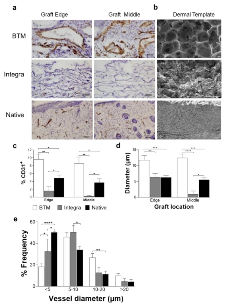
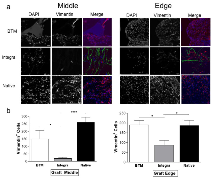
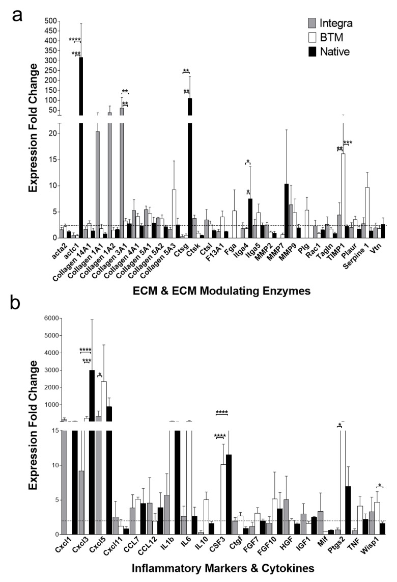
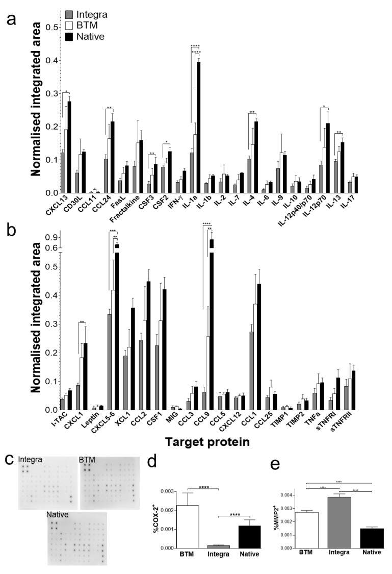
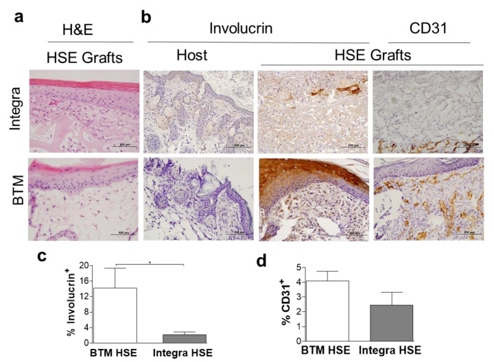
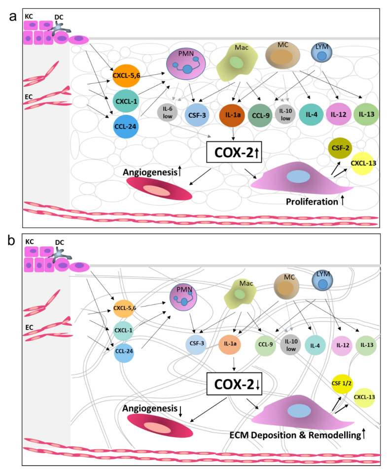
Engineered dermal templates have revolutionised the repair and reconstruction of skin defects. Their interaction with the wound microenvironment and linked molecular mediators of wound repair is still not clear. This study investigated the wound bed and acellular "off the shelf" dermal template interaction in a mouse model. Full-thickness wounds in nude mice were grafted with allogenic skin, and either collagen-based or fully synthetic dermal templates. Changes in the wound bed showed significantly higher vascularisation and fibroblast infiltration in synthetic grafts when compared to collagen-based grafts (P ≤ 0.05). Greater tissue growth was associated with higher prostaglandin-endoperoxide synthase 2 (Ptgs2) RNA and cyclooxygenase-2 (COX-2) protein levels in fully synthetic grafts. Collagen-based grafts had higher levels of collagen III and matrix metallopeptidase 2. To compare the capacity to form a double layer skin substitute, both templates were seeded with human fibroblasts and keratinocytes (so-called human skin equivalent or HSE). Mice were grafted with HSEs to test permanent wound closure with no further treatment required. We found the synthetic dermal template to have a significantly greater capacity to support human epidermal cells. In conclusion, the synthetic template showed advantages over the collagen-based template in a short-term mouse model of wound repair.

Keywords: COX-2; Integra®; NovoSorb® BTM; dermal templates; graft; human skin equivalent; inflammation; wound repair.


Conflict of interest statement

Authors have no conflict of interest to declare.

Figures

Figure 1
Figure 1
Detection of endothelial cells in mouse grafts. (a) CD31 positive endothelial cells were detected in grafts using IHC staining. Representative images of BTM, Integra®, and allogenic native skin grafts are presented (scale bar 50 µm). (b) SEM images of BTM, Integra®, and native mouse skin (scale bar 100 µm). (c) CD31 staining on graft edge and the middle was quantified and normalised for image area. CD31 staining was used to score vessel diameter (d), and the frequency of different vessel diameters (e). Values represent mean +/- SEM in each group (n = 4 mice per group) and analysed using unpaired t-test. * = p ≤ 0.05, ** = p ≤ 0.01, *** = p ≤ 0.001, **** = p ≤ 0.0001.
Figure 2
Figure 2
Mesenchymal host infiltration in mouse grafts. (a) Fibroblasts were detected by confocal microscopy using a vimentin antibody (in red) and DAPI (in blue). Dermal template autofluorescence is shown in green (scale bar 50 µm). (b) Vimentin positive cells were quantified on NIS Analysis software (Nikon, Japan) in six fields of view (n = 4–6 mice per group). Values represent mean +/− SEM in each group and analysed using unpaired t-test. * = p ≤ 0.05, **** = p < 0.0001.
Figure 3
Figure 3
RNA expression profiling (mouse wound healing RT2 profiler PCR array) of 2-week-long grafts. (a) Upregulated ECM structural and modifying enzyme genes identified in grafts by Ct comparison with host RNA. (b) Upregulated inflammation markers, including cytokines and chemokines. Selected targets produced an average of >2-fold change in at least one of the studied groups. Mean and SEM values presented for each group (n = 3 per group). * = p < 0.05, ** = p < 0.01, *** = p < 0.001, **** = p < 0.0001). A heatmap of all arrayed genes is available in Supplementary Data Figure S2.
Figure 4
Figure 4
Protein expression profiling of 2-week-long grafts. (a,b) Protein signals recorded for each target using 10-second exposure and normalised to positive controls. Mean and SEM values presented for each target (n = 4 per group) (a) Targets from rows 1 and 2 position F6 to rows 5 and 6 position B2 of the array). (b) Targets from rows 5 and 6 position B3 to rows 7 and 8 position I9 of the array. The full map of the array is provided in Supplementary Data Table S1. (c) Representative chemiluminescent protein array blots for each group. (d) Quantification of immunoperoxidase staining for inflammation marker, COX-2 (n = 4–8 mice per group), using unpaired t-test * = p < 0.05, ** = p < 0.01, *** = p < 0.001, **** = p < 0.0001. (e) Quantification of immunoperoxidase staining for ECM remodelling enzyme, MMP-2 (n = 4–8 mice per group). Results analysed using unpaired t-test, significant p-values as for panel c. Abbreviations: CCL1 (TCA-3), chemokine CC motif ligand 1; CCL2 (MCP-1), chemokine CC motif ligand 2; CCL3 (MIP-1α), chemokine CC motif ligand 3; CCL5 (RANTES), chemokine CC motif ligand 5; CCL9 (MIP1γ), chemokine CC motif ligand 9; CCL11 (Eotaxin), chemokine CC motif ligand 11; CCL24 (Eotaxin-2), chemokine CC motif ligand 24; CCL25 (TECK), chemokine CC motif ligand 25; CSF1 (M-CSF), colony-stimulating factor 1; CSF2 (GM-CSF), colony stimulating factor 2; CSF3 (G-CSF), colony stimulating factor 3; CXCL1 (KC), chemokine CXC motif ligand 1; CXCL5-6 (LIX), chemokine CXC motif ligand 5-6; CXCL12 (SDF-1), chemokine CXC motif ligand 12; CXCL13 (BLC), chemokine CXC motif ligand 13; IL1 or 6 or 10, interleukin 1 or 6 or 10; TIMP, tissue inhibitors of metalloproteinases; TNF, tumour necrosis factor, XCL1, lymphotactin α.
Figure 5
Figure 5
Neo-epidermis detection in 2-week-long HSE grafts. (a) Representative H&E staining of HSE constructed using Integra® (single layer) or BTM (single layer) two weeks post grafting (scale bar 100 µm). (b) Representative images of human-specific involucrin staining of mouse host and HSE grafts and CD31 in vivo (scale bar 100 µm). (c) Human involucrin staining of HSE grafts was quantified and Mean and SEM values are presented (n = 5–6 per group, *p value < 0.05). (d) CD31 expression was also quantified in HSE grafts. Mean and SEM values are presented (n = 5–6 per group).
Figure 6
Figure 6
A schematic diagram representing the key differences between Integra, BTM and allogenic native skin grafts in terms of inflammatory and wound healing mediators’ expression levels. Growth factors showing differential expression at protein levels in grafts and cell types known to secrete these growth factors are presented. (a) Allogenic native skin graft (and to a lesser degree BTM graft) triggers significantly higher inflammation response in the host, compared to (b) Integra®, resulting in greater fibroblast growth and vascularisation. Integra® grafts, however, are more progressed towards ECM remodelling phase within this time point, possibly triggered by an excessive amount of collagen in the graft. The arrows suggest pathways, based on the literature below, which link the growth factors to the grafting outcomes. Only the feed-forward pathways (and no inhibitory pathways) are presented. Abbreviations: polymorphonuclear cells (PMN), macrophages (Mac), mast cells (MC), basal keratinocytes (KC), endothelial cells (EC), and dendritic cells (DC) [17,19,21,22,23,24,25,26,27,28,29,30,31].

References

    1. Greenwood J.E., Dearman B.L. Split skin graft application over an integrating, biodegradable temporizing polymer matrix: Immediate and delayed. J. Burn Care Res. Off. Publ. Am. Burn Assoc. 2012;33:7–19. doi: 10.1097/BCR.0b013e3182372be9. - DOI - PubMed
    1. Burke J.F., Yannas I.V., Quinby W.C., Jr., Bondoc C.C., Jung W.K. Successful use of a physiologically acceptable artificial skin in the treatment of extensive burn injury. Ann. Surg. 1981;194:413–428. doi: 10.1097/00000658-198110000-00005. - DOI - PMC - PubMed
    1. Suzuki S., Matsuda K., Maruguchi T., Nishimura Y., Ikada Y. Further applications of “bilayer artificial skin”. Br. J. Plast. Surg. 1995;48:222–229. doi: 10.1016/0007-1226(95)90006-3. - DOI - PubMed
    1. Rygaard J. Skin grafts in nude mice. 1. Allografts in nude mice of three genetic backgrounds (BALB-c, C3H, C57-BL) Acta Pathol. Microbiol. Scand. A. 1974;82:80–92. - PubMed
    1. Landen N.X., Li D., Stahle M. Transition from inflammation to proliferation: A critical step during wound healing. Cell. Mol. Life Sci. 2016;73:3861–3885. doi: 10.1007/s00018-016-2268-0. - DOI - PMC - PubMed

Publication types

Grants and funding