. 2020 Jun 25;21(12):4520.
doi: 10.3390/ijms21124520.

The Presence of Seminal Plasma during Liquid Storage of Pig Spermatozoa at 17 °C Modulates Their Ability to Elicit In Vitro Capacitation and Trigger Acrosomal Exocytosis

Affiliations

The Presence of Seminal Plasma during Liquid Storage of Pig Spermatozoa at 17 °C Modulates Their Ability to Elicit In Vitro Capacitation and Trigger Acrosomal Exocytosis

Ana Paula Pinoti Pavaneli et al. Int J Mol Sci. .

Abstract

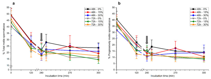
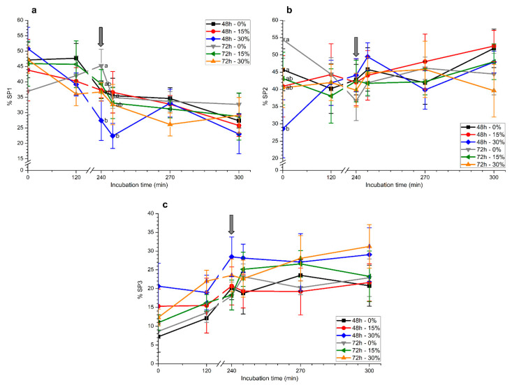
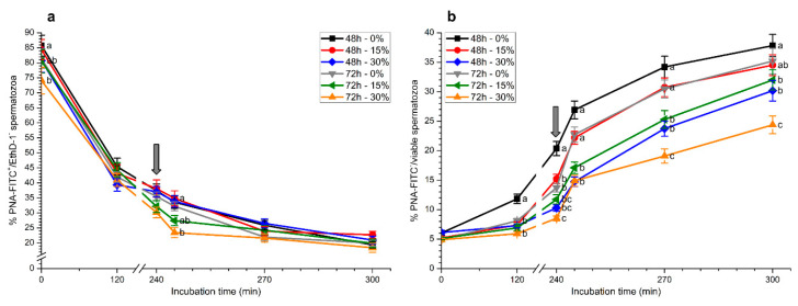
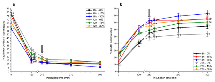
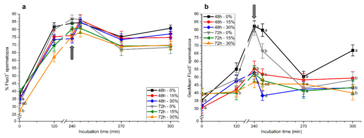
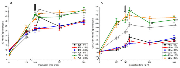
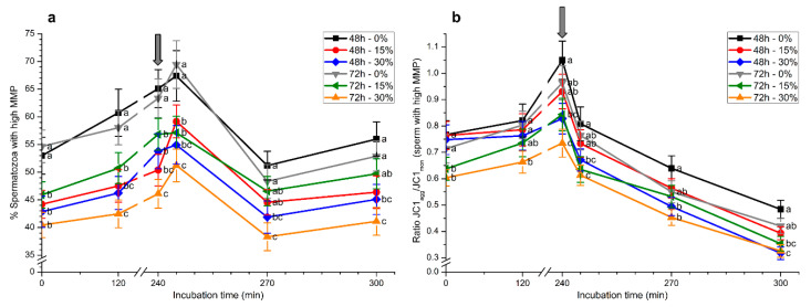
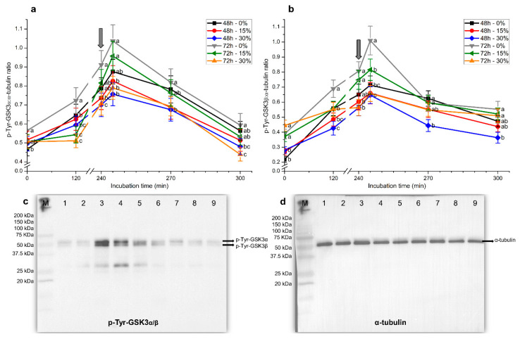
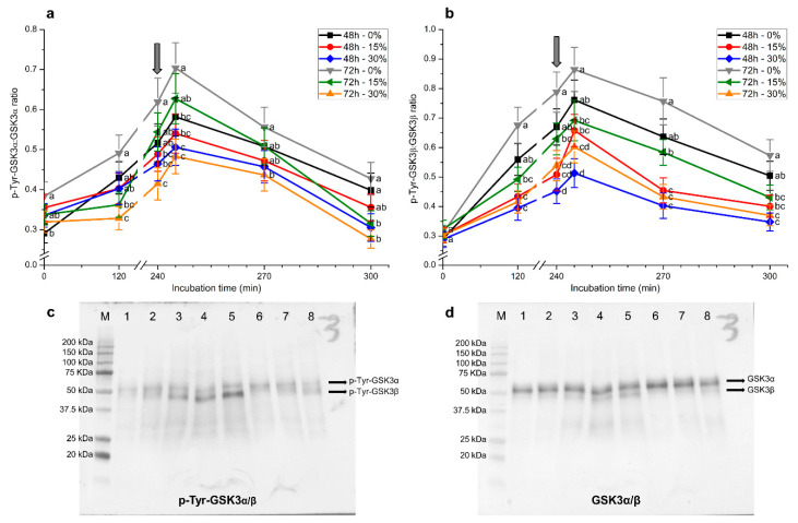
Although seminal plasma is essential to maintain sperm integrity and function, it is diluted/removed prior to liquid storage and cryopreservation in most mammalian species. This study sought to evaluate, using the pig as a model, whether storing semen in the presence of seminal plasma affects the sperm ability to elicit in vitro capacitation and acrosomal exocytosis. Upon collection, seminal plasma was separated from sperm samples, which were diluted in a commercial extender, added with seminal plasma (15% or 30%), and stored at 17 °C for 48 or 72 h. Sperm cells were subsequently exposed to capacitating medium for 4 h, and then added with progesterone to induce acrosomal exocytosis. Sperm motility, acrosome integrity, membrane lipid disorder, intracellular Ca2+ levels, mitochondrial activity, and tyrosine phosphorylation levels of glycogen synthase kinase-3 (GSK3)α/β were determined after 0, 2, and 4 h of incubation, and after 5, 30, and 60 min of progesterone addition. Results showed that storing sperm at 17 °C with 15% or 30% seminal plasma led to reduced percentages of viable spermatozoa exhibiting an exocytosed acrosome, mitochondrial membrane potential, intracellular Ca2+ levels stained by Fluo3, and tyrosine phosphorylation levels of GSK3α/β after in vitro capacitation and progesterone-induced acrosomal exocytosis. Therefore, the direct contact between spermatozoa and seminal plasma during liquid storage at 17 °C modulated their ability to elicit in vitro capacitation and undergo acrosomal exocytosis, via signal transduction pathways involving Ca2+ and Tyr phosphorylation of GSK3α/β. Further research is required to address whether such a modulating effect has any impact upon sperm fertilizing ability.

Keywords: acrosomal exocytosis; in vitro capacitation; seminal plasma; spermatozoa.


Conflict of interest statement

The authors declare that there is no conflict of interest that could be perceived as prejudicing the impartiality of the research reported herein.

Figures

Figure 1
Figure 1
Percentages of total (a) and progressively motile spermatozoa (b) during in vitro capacitation and progesterone-induced acrosomal exocytosis (300 min) after previous storage of spermatozoa at 17 °C with different concentrations of seminal plasma (0%, 15%, and 30%) for 48 h or 72 h. Grey arrow indicates the time at which 10 μg/mL progesterone was added to induce acrosomal exocytosis (i.e., 240 min). No significant differences (p > 0.05) between treatments were observed. Data are shown as mean ± standard error of the mean (SEM) for seven independent experiments.
Figure 2
Figure 2
Percentages of motile sperm populations ((a) SP1, (b) SP2, and (c) SP3) during in vitro capacitation and progesterone-induced acrosomal exocytosis after previous storage of spermatozoa at 17 °C with different concentrations of seminal plasma (0%, 15%, and 30%) for 48 h or 72 h. Grey arrow indicates the time at which 10 μg/mL progesterone was added to induce acrosomal exocytosis (i.e., 240 min). Different letters mean significant (p < 0.05) differences between treatments at a given time point. Data are shown as mean ± SEM for seven independent experiments.
Figure 3
Figure 3
Percentages of viable spermatozoa with an intact acrosome (PNA-FITC+/EthD-1; (a)) and with an exocytosed acrosome (PNA) in relation to total viable spermatozoa (b) during in vitro capacitation and progesterone-induced acrosomal exocytosis (300 min) after previous storage of spermatozoa at 17 °C with different concentrations of seminal plasma (0%, 15%, and 30%) for 48 h or 72 h. Grey arrow indicates the time at which 10 μg/mL progesterone was added to induce acrosomal exocytosis (i.e., 240 min). Different letters mean significant (p < 0.05) differences between treatments at a given time point. Data are shown as mean ± SEM for seven independent experiments.
Figure 4
Figure 4
Percentages of viable spermatozoa with low membrane lipid disorder (M540/YO-PRO-1; (a)) and of M540+ spermatozoa (b) during in vitro capacitation and progesterone-induced acrosomal exocytosis (300 min) after previous storage of spermatozoa at 17 °C with different concentrations of seminal plasma (0%, 15%, and 30%) for 48 h or 72 h. Grey arrow indicates the time at which 10 μg/mL progesterone was added to induce acrosomal exocytosis (i.e., 240 min). Different letters mean significant (p < 0.05) differences between treatments at a given time point. Data are shown as mean ± SEM for seven independent experiments.
Figure 5
Figure 5
Percentages (a) and geometric mean intensity (b) of Fluo3+ spermatozoa during in vitro capacitation and progesterone-induced acrosomal exocytosis (300 min) after previous storage of spermatozoa at 17 °C with different concentrations of seminal plasma (0%, 15%, and 30%) for 48 h or 72 h. Grey arrow indicates the time at which 10 μg/mL progesterone was added to induce acrosomal exocytosis (i.e., 240 min). Different letters mean significant (p < 0.05) differences between treatments at a given time point. Data are shown as mean ± SEM for seven independent experiments.
Figure 6
Figure 6
Percentages (a) and geometric mean intensity (b) of Rhod5+ spermatozoa during in vitro capacitation and progesterone-induced acrosomal exocytosis (300 min) after previous storage of spermatozoa at 17 °C with different concentrations of seminal plasma (0%, 15%, and 30%) for 48 h or 72 h. Grey arrow indicates the time at which 10 μg/mL progesterone was added to induce acrosomal exocytosis (i.e., 240 min). Different letters mean significant (p < 0.05) differences between treatments at a given time point. Data are shown as mean ± SEM for seven independent experiments.
Figure 7
Figure 7
Percentages of spermatozoa with high mitochondrial membrane potential (MMP; (a)) and their JC1agg/JC1mon ratios (b) during in vitro capacitation and progesterone-induced acrosomal exocytosis (300 min) after previous storage of spermatozoa at 17 °C with different concentrations of seminal plasma (0%, 15%, and 30%) for 48 h or 72 h. Grey arrow indicates the time at which 10 μg/mL progesterone was added to induce acrosomal exocytosis (i.e., 240 min). Different letters mean significant (p < 0.05) differences between treatments at a given time point. Data are shown as mean ± SEM for seven independent experiments.
Figure 8
Figure 8
Relative tyrosine phosphorylation levels (using α-tubulin as a loading control) for glycogen synthase kinase-3 (GSK3)α (a) and GSK3β (b) during in vitro capacitation and progesterone-induced acrosomal exocytosis (300 min) after previous storage of spermatozoa at 17 °C with different concentrations of seminal plasma (0%, 15%, and 30%) for 48 h or 72 h. Grey arrow indicates the time at which 10 μg/mL progesterone was added to induce acrosomal exocytosis (i.e., 240 min). Results are shown as mean ± SEM for seven separate experiments. Different letters mean significant (p < 0.05) differences between treatments at a given time point. Representative blots for p-Tyr-GSK3α/β (c) and α-tubulin (d). Lanes: (M) protein ladder; (1) 72 h, 0% SP, 0 min; (2) 72 h, 0% SP, 120 min; (3) 72 h, 0% SP, 240 min; (4) 72 h, 0% SP, 245 min; (5) 72 h, 0% SP, 270 min; (6) 72 h, 0% SP, 300 min; (7) 72 h, 15% SP, 0 min; (8) 72 h, 15% SP, 120 min; (9) 72 h, 15% SP, 240 min.
Figure 9
Figure 9
Relative tyrosine phosphorylation levels (using total GSK3α/β as a loading control) for GSK3α (a) and GSK3β (b) during in vitro capacitation and progesterone-induced acrosomal exocytosis (300 min) after previous storage of spermatozoa at 17 °C with different concentrations of seminal plasma (0%, 15%, and 30%) for 48 h or 72 h. Grey arrow indicates the time at which 10 μg/mL progesterone was added to induce acrosomal exocytosis (i.e., 240 min). Results are shown as mean ± SEM for seven separate experiments. Different letters mean significant (p < 0.05) differences between treatments at a given time point. Representative blots for p-Tyr-GSK3α/β (c) and α-tubulin (d). Lanes: (M) protein ladder; (1) 72 h, 0% SP, 0 min; (2) 72 h, 0% SP, 120 min; (3) 72 h, 0% SP, 240 min; (4) 72 h, 0% SP, 245 min; (5) 72 h, 0% SP, 270 min; (6) 72 h, 0% SP, 300 min; (7) 72 h, 15% SP, 0 min; (8) 72 h, 15% SP, 120 min.

Similar articles

Cited by

References

    1. Juyena N.S., Stelletta C. Seminal plasma: An essential attribute to spermatozoa. J. Androl. 2012;33:536–551. doi: 10.2164/jandrol.110.012583. - DOI - PubMed
    1. Barranco I., Padilla L., Parrilla I., Álvarez-Barrientos A., Pérez-Patiño C., Peña F.J., Martínez E.A., Rodriguez-Martínez H., Roca J. Extracellular vesicles isolated from porcine seminal plasma exhibit different tetraspanin expression profiles. Sci. Rep. 2019;9:11584. doi: 10.1038/s41598-019-48095-3. - DOI - PMC - PubMed
    1. Alvarez-Rodriguez M., Ljunggren S.A., Karlsson H., Rodriguez-Martinez H. Exosomes in specific fractions of the boar ejaculate contain CD44: A marker for epididymosomes? Theriogenology. 2019;140:143–152. doi: 10.1016/j.theriogenology.2019.08.023. - DOI - PubMed
    1. Du J., Shen J., Wang Y., Pan C., Pang W., Diao H., Dong W. Boar seminal plasma exosomes maintain sperm function by infiltrating into the sperm membrane. Oncotarget. 2016;7:58832–58847. doi: 10.18632/oncotarget.11315. - DOI - PMC - PubMed
    1. Bai R., Latifi Z., Kusama K., Nakamura K., Shimada M., Imakawa K. Induction of immune-related gene expression by seminal exosomes in the porcine endometrium. Biochem. Biophys. Res. Commun. 2018;495:1094–1101. doi: 10.1016/j.bbrc.2017.11.100. - DOI - PubMed

MeSH terms

LinkOut - more resources