Review
. 2020 Jun 25;13(12):2853.
doi: 10.3390/ma13122853.

Hydrogel Dressings for the Treatment of Burn Wounds: An Up-To-Date Overview

Affiliations
Review

Hydrogel Dressings for the Treatment of Burn Wounds: An Up-To-Date Overview

Alexandra Elena Stoica et al. Materials (Basel). .

Abstract

Globally, the fourth most prevalent devastating form of trauma are burn injuries. Ideal burn wound dressings are fundamental to facilitate the wound healing process and decrease pain in lower time intervals. Conventional dry dressing treatments, such as those using absorbent gauze and/or absorbent cotton, possess limited therapeutic effects and require repeated dressing changes, which further aggravate patients' suffering. Contrariwise, hydrogels represent a promising alternative to improve healing by assuring a moisture balance at the burn site. Most studies consider hydrogels as ideal candidate materials for the synthesis of wound dressings because they exhibit a three-dimensional (3D) structure, which mimics the natural extracellular matrix (ECM) of skin in regard to the high-water amount, which assures a moist environment to the wound. There is a wide variety of polymers that have been used, either alone or blended, for the fabrication of hydrogels designed for biomedical applications focusing on treating burn injuries. The aim of this paper is to provide an up-to-date overview of hydrogels applied in burn wound dressings.

Keywords: burn injury; hydrogels; skin regeneration; wound dressing; wound healing.


Conflict of interest statement

The authors declare no conflict of interest.

Figures

Figure 1
Figure 1
Different depth of invasion for burn injury [13]. Reprinted from an open-access source.
Figure 2
Figure 2
Structure of different types of dressings [24]. Reprinted from an open-access source.
Figure 3
Figure 3
Properties of an ideal wound dressing [19]. Reprinted from an open-access source.
Figure 4
Figure 4
Healing of deep second-degree burn of rat skin with different treatments. (A): Photographs of deep second burn wounds at 0, 7, 14, 21 and 28 days. (B): Wound healing rate with different treatments. Positive control group, treated with commercial product (3MTM TegadermTM hydrocolloid dressing); Collagen hydrogels group, treated with collagen hydrogel dressing containing 10 mg/mL PSC; Blank control group, without any treatment after wound burned [90]. Reprinted from an open-access source.
Figure 5
Figure 5
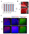
Characterization of the cytocompatibility of the hydrogels. (A) MTT assay of human fibroblast cells grown in the presence of different hydrogels. Wells treated with ethanol were used as positive controls. n.s: no statistically significant groups. The data are shown as means ± standard deviations (n = 3). (B) Representative SEM images of fibroblast cell adhesion and proliferation on the surface of the 1%_XG/KGM_(60/40) hydrogel, after 24 h and 72 h of incubation. (C) Confocal laser scanning microscopy (CLSM) images of cell internalization in 1%_XG/KGM_(60/40) after 24 h and 72 h, where different colors correspond to distinct depth values (as indicated in the color-coding scale) [95]. Reprinted from an open-access source.
Figure 6
Figure 6
Macroscopic images of the hydrogels XG/KGM [95]. Reprinted from an open-access source.

References

    1. Koehler J., Brandl F.P., Goepferich A.M. Hydrogel wound dressings for bioactive treatment of acute and chronic wounds. Eur. Polym. J. 2018;100:1–11. doi: 10.1016/j.eurpolymj.2017.12.046. - DOI
    1. Dong Y., Cui M., Qu J., Wang X., Kwon S.H., Barrera J., Elvassore N., Gurtner G.C. Conformable hyaluronic acid hydrogel delivers adipose-derived stem cells and promotes regeneration of burn injury. Acta Biomater. 2020;108:56–66. doi: 10.1016/j.actbio.2020.03.040. - DOI - PubMed
    1. Jeschke M.G., Gauglitz G.G. Pathophysiology of Burn Injuries. In: Jeschke M.G., Kamolz L.-P., Sjöberg F., Wolf S.E., editors. Handbook of Burns Volume 1. Springer; Berlin/Heidelberg, Germany: 2020. pp. 229–245.
    1. Kaddoura I., Abu-Sittah G., Ibrahim A., Karamanoukian R., Papazian N. Burn injury: Review of pathophysiology and therapeutic modalities in major burns. Ann. Burns Fire Disasters. 2017;30:95–102. - PMC - PubMed
    1. Brassolatti P., de Andrade A.L.M., Bossini P.S., Otterço A.N., Parizotto N.A. Evaluation of the low-level laser therapy application parameters for skin burn treatment in experimental model: A systematic review. Lasers Med. Sci. 2018;33:1159–1169. doi: 10.1007/s10103-018-2526-5. - DOI - PubMed

LinkOut - more resources