Review
. 2020 Jul 2;21(13):4724.
doi: 10.3390/ijms21134724.

Stimuli-Responsive Materials for Tissue Engineering and Drug Delivery

Affiliations
Review

Stimuli-Responsive Materials for Tissue Engineering and Drug Delivery

Sofia Municoy et al. Int J Mol Sci. .

Abstract

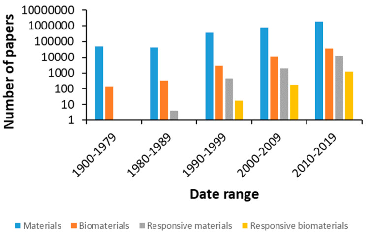
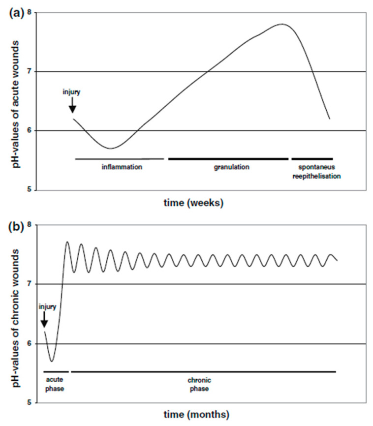
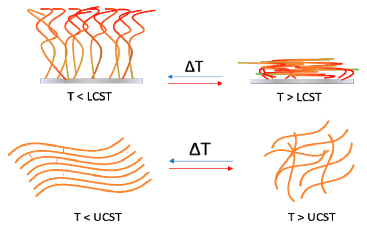
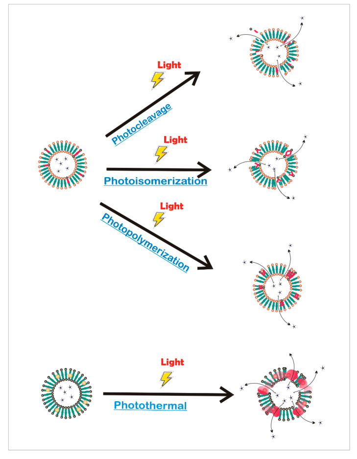
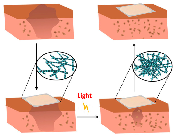
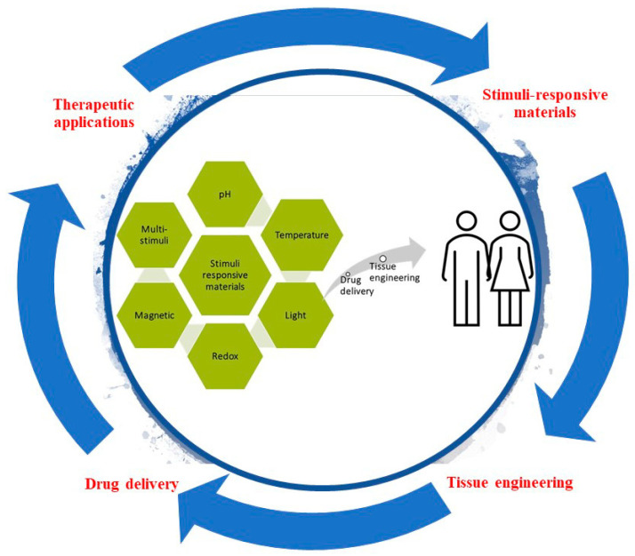
Smart or stimuli-responsive materials are an emerging class of materials used for tissue engineering and drug delivery. A variety of stimuli (including temperature, pH, redox-state, light, and magnet fields) are being investigated for their potential to change a material's properties, interactions, structure, and/or dimensions. The specificity of stimuli response, and ability to respond to endogenous cues inherently present in living systems provide possibilities to develop novel tissue engineering and drug delivery strategies (for example materials composed of stimuli responsive polymers that self-assemble or undergo phase transitions or morphology transformations). Herein, smart materials as controlled drug release vehicles for tissue engineering are described, highlighting their potential for the delivery of precise quantities of drugs at specific locations and times promoting the controlled repair or remodeling of tissues.

Keywords: biomaterials; drug delivery; light-responsive; pH-responsive; redox-responsive; stimuli-responsive materials; thermoresponsive; tissue engineering.


Conflict of interest statement

The authors declare no conflict of interest.

Figures

Figure 1
Figure 1
Number of publications on related topics in the Web of Science database with respect to time.
Figure 2
Figure 2
(a) Course of pH milieu in acute wounds. (b) Course of pH milieu in chronic wounds. Reprinted by permission from Springer Nature, Arch Dermatol Res, in [57], Copyright (2006).
Figure 3
Figure 3
In vitro competitive co-culture MC3T3-E1/S. aureus after 6 h, 24 h and 3 days of incubation onto MGHA and MGHA-Levo 3D scaffolds. Material refraction in green, preostoblastic nuclei and bacteria in blue (DAPI) and actin-fibrous of preosteoblast cytoplasm in red (phalloidin). Reprinted from reference [61], Copyright (2018), with permission from Elsevier.
Figure 4
Figure 4
Representative in vivo (A) and ex vivo (B) images of 4T1 tumor-bearing BALB/C mice 24 h after injection of Cy5 labeled antagomir-10b-loaded liposomes. The black arrows in (A) indicated the location of tumors. Reprinted from [65], Copyright (2015), with permission from Elsevier.
Figure 5
Figure 5
Representative scheme of PNIPAAm gelation at temperatures above its lower critical solution temperature (LCST) (top) and below its upper critical solution temperature (UCST) (bottom).
Figure 6
Figure 6
Osteoinductive and osteoconductive activities of the GO-P hybrid scaffold in vitro. (A) AdGFP or AdBMP9-infected iMADs were mixed with PPCNg (a) or GO-P (b) and examined at 48 h after infection under bright field (BF) or GFP fluorescence microscope (GFP). Representative images are shown. (B,C) ALP activity analysis. AdGFP or AdBMP9-transduced iMADs were mixed with PPCNg or GO-P and seeded in 24-well plates. ALP staining was carried out on day 5 (B), while quantitative ALP assay was conducted at 3, 5, and 7 days after infection (C). All assays were done in triplicate. * p < 0.05 and ** p < 0.01 when compared to respective GFP groups. Reproduced from [73]. Copyright © 2018 American Chemical Society.
Figure 7
Figure 7
Release from liposomes controlled by photochemical reactions. Photomediated release can be achieved by photocleavage, photoisomerization, photopolymerization, and photothermal reactions.
Figure 8
Figure 8
Scheme showing a photoreactive composite for tissue regeneration. Photopolymerization allows to initiate and propagate a polymerization of networks in situ, creating covalently crosslinked hydrogels for tissue reconstruction.
Figure 9
Figure 9
(a) An iterative synthesis was used to prepare each even-numbered (n = 2, 4, 6, 8, and 10) oligoviologen by (i) alternating between the excessive addition (20 equiv) of tosyl end-capped hexaethylene glycol (HEG-Tos) and 4,4′-bipyridine (BIPY) in MeCN at 130 °C for 12–16 h in a closed reaction vessel. (b) The synthetic cycle begins (green box) with a BIPY end-capped HEG (HEG-BIPY2+), and the oligomer is grown iteratively, with only intermittent precipitations in MeCN:PhMe, followed by centrifugation in order to isolate each product. At any point in the cycle, the BIPY end-capped precursor can be removed and (ii) functionalized with terminal azide groups (red box) through the excessive addition (35 equiv, MeCN, 130 °C, 20 h) of a tosylated diethylene glycol possessing one azide at its terminus (Tos-DEG-N3). (c) Synthesis of the click-based hydrogel involves 2 equiv of bis-azide-terminated linkers, where 95 mol % of the 2 equiv is composed of polyethylene glycol (PEG-N3) and 5 mol % consists of the oligoviologen (nV(2n) +-N3), to 1 equiv of the tetra-alkyne cross-linker (TAXL). Published in [176]. Copyright© 2017 American Chemical Society.
Figure 10
Figure 10
Schematic illustration of the internal structure of the actuator as a result of actuation. The creation of dimensional gradients within the polymer layer was responsible for the fast and efficient electromechanical deformation of the actuator. Reprinted with permission from [194]. Copyright (2013) Nature Publishing Group.
Figure 11
Figure 11
Proposed illustration of one-step ligand exchange reaction, ligand candidates, and the target solvents. Reprinted (adapted) with permission from [214]. Copyright (2014) American Chemical Society.
Figure 12
Figure 12
Schematic illustration of therapeutic applications and stimuli responsive materials.

References

    1. Lumelsky N., O’Hayre M., Chander P., Shum L., Somerman M.J. Autotherapies: Enhancing Endogenous Healing and Regeneration. Trends Mol. Med. 2018;24:919–930. doi: 10.1016/j.molmed.2018.08.004. - DOI - PubMed
    1. Lavrador P., Gaspar V.M., Mano J.F. Stimuli-responsive nanocarriers for delivery of bone therapeutics–Barriers and progresses. J. Control. Release. 2018;273:51–67. doi: 10.1016/j.jconrel.2018.01.021. - DOI - PMC - PubMed
    1. Rogina A., Ressler A., Matić I., Gallego Ferrer G., Marijanović I., Ivanković M., Ivanković H. Cellular hydrogels based on pH-responsive chitosan-hydroxyapatite system. Carbohydr. Polym. 2017;166:173–182. doi: 10.1016/j.carbpol.2017.02.105. - DOI - PubMed
    1. Echazú M.I.A., Olivetti C.E., Peralta I., Alonso M.R., Anesini C., Perez C.J., Alvarez G.S., Desimone M.F. Development of pH-responsive biopolymer-silica composites loaded with Larrea divaricata Cav. extract with antioxidant activity. Colloids Surfaces B Biointerfaces. 2018;169:82–91. doi: 10.1016/j.colsurfb.2018.05.015. - DOI - PubMed
    1. Parani M., Lokhande G., Singh A., Gaharwar A.K. Engineered Nanomaterials for Infection Control and Healing Acute and Chronic Wounds. ACS Appl. Mater. Interfaces. 2016;8:10049–10069. doi: 10.1021/acsami.6b00291. - DOI - PubMed

MeSH terms

LinkOut - more resources