. 2020 Jul 2;9(3):29.
doi: 10.3390/antib9030029.

Rapid Isolation of Rare Isotype-Switched Hybridoma Variants: Application to the Generation of IgG2a and IgG2b MAb to CD63, a Late Endosome and Exosome Marker

Affiliations

Rapid Isolation of Rare Isotype-Switched Hybridoma Variants: Application to the Generation of IgG2a and IgG2b MAb to CD63, a Late Endosome and Exosome Marker

Stéphanie Charrin et al. Antibodies (Basel). .

Abstract

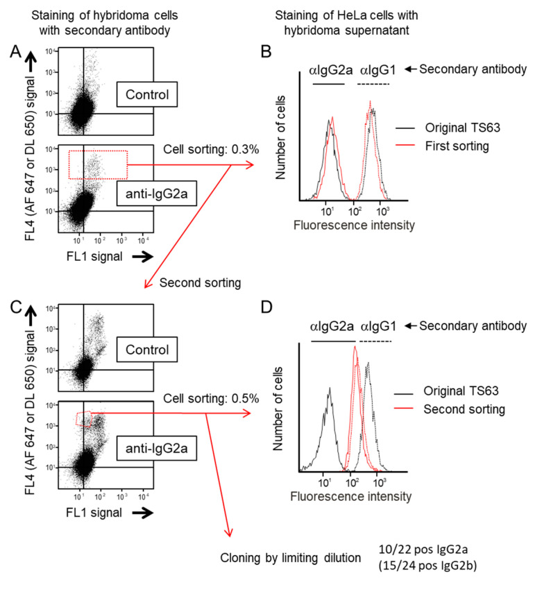
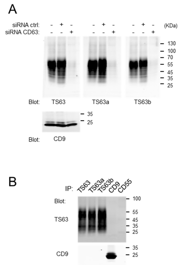
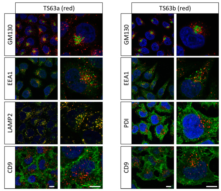
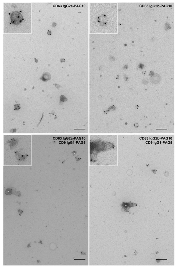
CD63, a member of the tetraspanin superfamily, is used as a marker of late endosomes and lysosome-related organelles, as well as a marker of exosomes. Here, we selected rare isotype variants of TS63 by sorting hybridoma cells on the basis of their high expression of surface immunoglobulins of the IgG2a and IgG2b subclass. Pure populations of cells secreting IgG2a and IgG2b variants of TS63 (referred to as TS63a and TS63b) were obtained using two rounds of cell sorting and one limited dilution cloning step. We validate that these new TS63 variants are suitable for co-labeling with mAb of the IgG1 subclass directed to other molecules, using anti mouse subclass antibodies, and for the labeling of exosomes through direct binding to protein A-coated gold particles. These mAbs will be useful to study the intracellular localization of various proteins and facilitate electron microscopy analysis of CD63 localization.

Keywords: CD63; exosomes; isotype switch; late endosomes.


Conflict of interest statement

The mAb TS63 was produced in our laboratory and is licensed for reselling. The funders had no role in the design of the study; in the collection, analyses, or interpretation of data; in the writing of the manuscript, or in the decision to publish the results.

Figures

Figure 1
Figure 1
Selection of IgG2a variants of TS63. (A) Hybridoma cells were labelled with either a control secondary antibody (a goat anti-rabbit coupled to Dylight 650 (DL 650)) or a goat anti-mouse IgG2a coupled to Alexa Fluor 647 (AF 647). The dot plots show for each cell the value of the Dylight 650 or Alexa 647 fluorescence in the far red Channel (FL4) and that of the autofluorescence in the green channel (FL1). Note that there is no difference between the two labelings indicating that most of the “positive” signal is non-specific. The gate used to sort the cells with the highest level of staining is drawn in the bottom dot-blot. (B) After being grown for a few days, the supernatant of the sorted cells was used to stain HeLa cells by indirect immunofluorescence, using either anti (α)-IgG1 or anti-IgG2a polyclonal antibodies coupled to Alexa 647 as secondary reagents. A staining with the conditioned medium of parental TS63 cells was performed in parallel. The fluorescence staining of the cells was analyzed by flow-cytometry. Note that the supernatant of sorted cells stains HeLa cells slightly better than that of parental cells when the binding of the mAb is revealed by an anti-IgG2a antibody. (C) After amplification, the cells sorted in A were subjected to a second sorting. Note that there is a specific cell population uniquely detected by the anti-mouse IgG2a labeling. The gate used to sort the cells is drawn in the bottom dot-blot. (D) Labelling of HeLa cells as in B after growing the cells sorted the second time for a few days. Note that the staining of HeLa cells by the supernatant of the sorted cells is similar whether the binding of the mAb is revealed by an anti-IgG2a antibody or an anti-IgG1 antibody.
Figure 2
Figure 2
Validation that the TS63 variants recognize CD63. (A) Western-blot analysis, using the different TS63 variant or a CD9 mAb as a control, of HeLa cell lysates after treatment or not with a siRNA targeting CD63 or a control siRNA. (B) Immunoprecipitations using the different TS63 variants or CD9 and CD55 mAb as a control. The presence of CD63 and CD9 in the immunoprecipitates was analyzed by Western-blot using biotin-labeled TS63 and TS9 mAbs.
Figure 3
Figure 3
Confocal microscopy analysis of CD63 distribution and comparison with other organelle markers. HeLa cells were grown for two days on coverslips, fixed, permeabilized and stained with a combination of either TS63a or TS63b, and mAbs of different subclasses to markers of different compartments. The binding of the antibodies was revealed using goat polyclonal antibodies to mouse Ig subclasses coupled to AlexaFluor 488 or AlexaFluor 568. GM130: cis-Golgi; EEA1, early endosome; LAMP2: late endosome; PDI: endoplasmic reticulum; CD9: plasma membrane. Bar: 10 µm.
Figure 4
Figure 4
Immunogold labelling of extracellular vesicles with TS63a and TS63b.Top panels: TS63a or TS63b were used to label extracellular vesicles derived from MNT-1 cells. Both antibodies revealed specific and efficient labelling of a proportion of extracellular vesicles. Bottom panels: TS63a or TS63b were used to co-label extracellular vesicles derived from MNT-1 cells with anti-CD9 antibody. Both antibodies displayed distinct and specific labelling for each tetraspanin (10 nm gold particles for CD63 and 5 nm particles for CD9) and revealed subpopulations of extracellular singly labelled for either CD63 or CD9 (indicated by white star) or co-labelled for both. Inset shows a zoom-in of each panel. Scale bar: 200 nm.

References

    1. Charrin S., Jouannet S., Boucheix C., Rubinstein E. Tetraspanins at a glance. J. Cell Sci. 2014;127:3641–3648. doi: 10.1242/jcs.154906. - DOI - PubMed
    1. Pols M.S., Klumperman J. Trafficking and function of the tetraspanin CD63. Exp. Cell Res. 2009;315:1584–1592. doi: 10.1016/j.yexcr.2008.09.020. - DOI - PubMed
    1. Yoshida T., Kawano Y., Sato K., Ando Y., Aoki J., Miura Y., Komano J., Tanaka Y., Koyanagi Y. A CD63 mutant inhibits T-cell tropic human immunodeficiency virus type 1 entry by disrupting CXCR4 trafficking to the plasma membrane. Traffic. 2008;9:540–558. doi: 10.1111/j.1600-0854.2007.00700.x. - DOI - PubMed
    1. Duffield A., Kamsteeg E.J., Brown A.N., Pagel P., Caplan M.J. The tetraspanin CD63 enhances the internalization of the H,K-ATPase beta-subunit. Proc. Natl. Acad. Sci. USA. 2003;100:15560–15565. doi: 10.1073/pnas.2536699100. - DOI - PMC - PubMed
    1. Edgar J.R., Eden E.R., Futter C.E. Hrs- and CD63-dependent competing mechanisms make different sized endosomal intraluminal vesicles. Traffic. 2014;15:197–211. doi: 10.1111/tra.12139. - DOI - PMC - PubMed

LinkOut - more resources