

MicroRNA Milk Exosomes: From Cellular Regulator to Genomic Marker
- PMID: 32630756
- PMCID: PMC7401532
- DOI: 10.3390/ani10071126
MicroRNA Milk Exosomes: From Cellular Regulator to Genomic Marker
Abstract
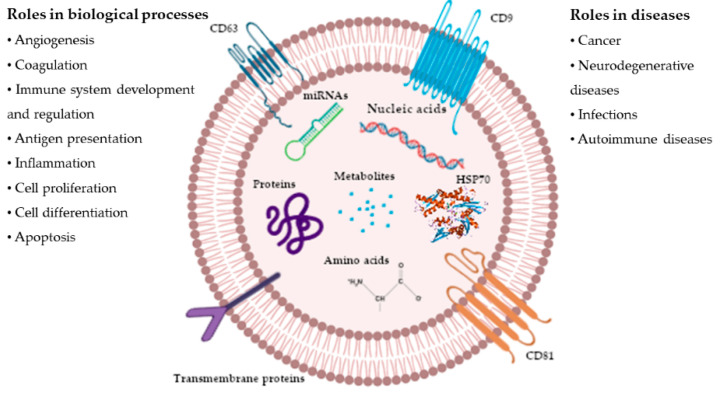
Recent advances in ruminants' milk-derived exosomes (EXO) have indicated a role of microRNAs (miRNAs) in cell-to-cell communication in dairy ruminants. The miRNAs EXO retain peculiar mechanisms of uptake from recipient cells, which enables the selective delivery of cargos, with a specific regulation of target genes. Although many studies have been published on the miRNAs contained in milk, less information is available on the role of miRNAs EXO, which are considered stable over time and resistant to digestion and milk processing. Several miRNAs EXO have been implicated in the cellular signaling pathway, as in the regulation of immune response. Moreover, they exert epigenetic control, as extenuating the expression of DNA methyltransferase 1. However, the study of miRNAs EXO is still challenging due to the difficulty of isolating EXO. In fact, there are not agreed protocols, and different methods, often time-consuming, are used, making it difficult to routinely process a large number of samples. The regulation of cell functions in mammary glands by miRNAs EXO, and their applications as genomic markers in livestock, is presented.
Keywords: bovine milk; exosomes; genome; mastitis; miRNA; ruminants.
Conflict of interest statement
The authors declare no conflict of interest. The funders had no role in the design of the study; in the collection, analyses, or interpretation of data; in the writing of the manuscript, or in the decision to publish the results.
Figures
References
-
- Sedykh S.E., Burkova E.E., Purvinsh L.V., Klemeshova D.A., Ryabchikova E.I., Nevinsky G.A. Milk exosomes: Isolation, biochemistry, morphology, and perspectives of use. In: De Bona A.G., Reales-Calderon J.A., editors. Extracellular Vesicles and Their Importance in Human Health. 1st ed. IntechOpen; London, UK: 2020. pp. 1–28.
-
- Rani S., O’Brien K., Kelleher F.C., Corcoran C., Germano S., Radomski M.W., Crown J., O’Driscoll L. Isolation of exosomes for subsequent mRNA, microRNA, and protein profiling. Methods Mol. Biol. 2011;784:181–195. - PubMed
-
- Lötvall J., Hill A.F., Hochberg F., Buzás E.I., Di Vizio D., Gardiner C., Gho Y.S., Kurochkin I.V., Mathivanan S., Quesenberry P., et al. Minimal experimental requirements for definition of extracellular vesicles and their functions: A position statement from the International Society for Extracellular Vesicles. J. Extracell. Vesicles. 2014;3:26913. doi: 10.3402/jev.v3.26913. - DOI - PMC - PubMed
Publication types
Grants and funding
LinkOut - more resources
Full Text Sources
