Review
. 2020 Jul 2;8(7):991.
doi: 10.3390/microorganisms8070991.

Middle East Respiratory Syndrome Coronavirus (MERS-CoV): State of the Science

Affiliations
Review

Middle East Respiratory Syndrome Coronavirus (MERS-CoV): State of the Science

Ahmed Mostafa et al. Microorganisms. .

Abstract

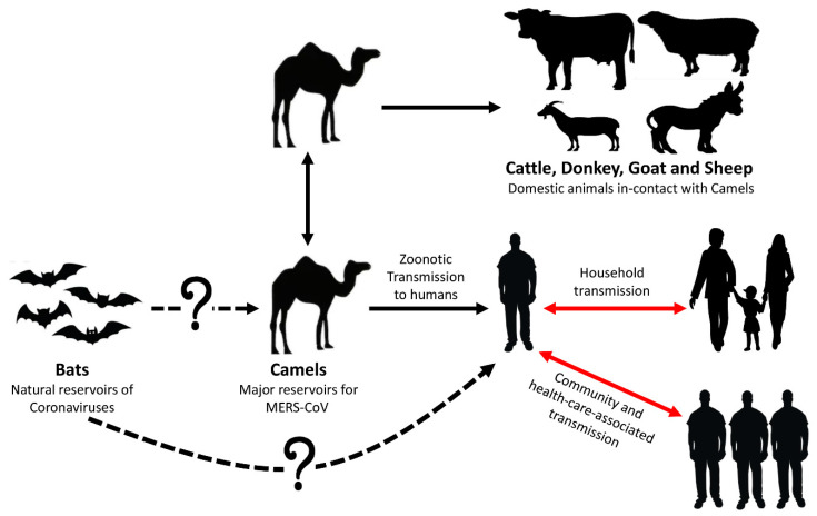
Coronaviruses belong to a large family of viruses that can cause disease outbreaks ranging from the common cold to acute respiratory syndrome. Since 2003, three zoonotic members of this family evolved to cross species barriers infecting humans and resulting in relatively high case fatality rates (CFR). Compared to Severe Acute Respiratory Syndrome Coronavirus (SARS-CoV, CFR = 10%) and pandemic Severe Acute Respiratory Syndrome Coronavirus 2 (SARS-CoV-2, CFR = 6%), the Middle East Respiratory Syndrome Coronavirus (MERS-CoV) has scored the highest CFR (approximately 35%). In this review, we systematically summarize the current state of scientific knowledge about MERS-CoV, including virology and origin, epidemiology, zoonotic mode of transmission, and potential therapeutic or prophylactic intervention modalities.

Keywords: MERS-CoV; coronavirus; epidemiology; zoonotic disease.


Conflict of interest statement

The authors declare no conflict of interest. The funders had no role in the design of the study; in the collection, analyses, or interpretation of data; in the writing of the manuscript, or in the decision to publish the results.

Figures

Figure 1
Figure 1
Zoonotic mode of transmission of MERS-CoV. The three routes of transmission (camel-to-camel, camel-to-human, and human-to-human) were confirmed. The prevalence of MERS-CoV in domestic animals in-contact with camels was recently identified [43]. As depicted by the dotted line, bat-to-camel and bat-to-human direct transmission of MERS-CoV have not been confirmed. Human-to-human transmission of the virus occurs after close contact with an infected case in households and healthcare settings (red arrows).
Figure 2
Figure 2
Three clades of MERS-CoV based on a rooted phylogenetic tree of 484 complete genomes of MERS-CoV strains from camel and human cases. MERS-CoV isolates are divided into three separate clades: A, B, and C. Clades A and B are prevalent in the Arabian Peninsula and other non-African world countries. Clade C is mainly circulating in African countries. The optimal tree with the sum of branch length = 0.11869958 is shown with scale bar = 0.0005 (5.0E−4).
Figure 3
Figure 3
Schematic diagram of the MERS-CoV genome and naturally selected aa substitutions in spike protein. (A) The genomic structure of MERS-CoV (30.1 kb in length), illustrating sub-genomic viral RNA transcripts. (B) Schematic structure of the MERS-CoV S protein and its functional domains, including the N-terminal domain (NTD), receptor-binding domain (RBD), receptor-binding motif (RBM), fusion peptide (FP), heptad repeat region 1 and 2 (HR1 and HR2), transmembrane region (TM), and cytoplasmic tail (CP). (C) Since the first documentation of MERS-CoV in 2012 in KSA, the virus circulated in camels and occasionally humans to naturally acquire distinct adaptive amino acid (aa) substitutions.
Figure 4
Figure 4
Schematic representation of Middle East respiratory syndrome coronavirus (MERS-CoV) replication cycle and key targets for antiviral activity. The spike protein of MERS-CoV initiates host cell infection via binding its receptor-binding domain (RBD) in the S1 subunit into cellular receptor dipeptidyl peptidase 4 (DPP4), originally known as the lymphocyte cell surface protein CD26. Following binding, the viral particle in the form of an endosome internalizes into the cytosol via acid-dependent proteolytic cleavage of S protein by a cathepsin or TMPRRS2. To release the viral genome (+ssRNA) into the cytoplasm, fusion of the viral and cellular membranes within acidified endosome occurs. Initially, the replicase gene, which encodes the largest two open-reading frames “ORF1a and ORF1ab”, is translated to express two co-terminal polyproteins, pp1a and pp1ab. These polyproteins are further cleaved by virus-encoded proteases “papain-like protease PLpro and 3C-like protease 3CLpro" into 16 mature nonstructural proteins (nsp) including viral polymerase subunits. Essential elements for viral genome replication are gathered as RNA replication–transcription complexes (RTCs) within the endoplasmic reticulum-derived double-membrane vesicles (DMVs). The RTCs drive the production of intermediate negative-sense viral genome (–ssRNA) transcript. During replication, the –ssRNA genome is used as a template for generating nascent +ssRNA. Along with the continuous transcription to generate the nascent full-length coding +ssRNA, sub-genomic RNAs, including those encoding all essential structural proteins (spike (S), envelope (E), membrane (M), and nucleocapsid (N)), are produced via discontinuous transcription. Nucleocapsid and nascent genomic RNA are assembled together in the cytoplasm and further transported into the lumen of the endoplasmic reticulum (ER)–Golgi intermediate compartment (ERGIC). Meanwhile, the S, E, and M sub-genomic RNAs are translated and inserted into the membrane of the rough endoplasmic reticulum (ER), from where they are transported to interact with RNA-encapsidated N proteins in the ERGIC, forming a mature viral particle. Via exocytosis, the nascent viral particle is then released from the infected cell. The repurposed therapeutic drugs undergoing preclinical and clinical trials against MERS-CoV in the context of host pathways and virus replication mechanisms are represented in the figure. The symbol ⊣ refers to inhibition.

References

    1. Gonzalez J.M., Gomez-Puertas P., Cavanagh D., Gorbalenya A.E., Enjuanes L. A comparative sequence analysis to revise the current taxonomy of the family Coronaviridae. Arch. Virol. 2003;148:2207–2235. doi: 10.1007/s00705-003-0162-1. - DOI - PMC - PubMed
    1. ICTV ICTV Taxonomy History for Coronavirinae Virus Taxonomy: 2011 Release. [(accessed on 20 August 2014)]; Available online: http://www.ictvonline.org/virusTaxonomy.asp?taxnode_id=20110624.
    1. Perlman S., Netland J. Coronaviruses post-SARS: Update on replication and pathogenesis. Nat. Rev. Microbiol. 2009;7:439–450. doi: 10.1038/nrmicro2147. - DOI - PMC - PubMed
    1. Zaki A.M., van Boheemen S., Bestebroer T.M., Osterhaus A.D., Fouchier R.A. Isolation of a novel coronavirus from a man with pneumonia in Saudi Arabia. N. Engl. J. Med. 2012;367:1814–1820. doi: 10.1056/NEJMoa1211721. - DOI - PubMed
    1. FAO MERS-CoV Situation Update. [(accessed on 30 June 2020)]; Available online: http://www.fao.org/ag/againfo/programmes/en/empres/mers/situation_update....

LinkOut - more resources