Review
. 2020 Jun 27;50(3):169-182.
doi: 10.4274/tjo.galenos.2019.60308.

Behçet's Uveitis: Current Diagnostic and Therapeutic Approach

Affiliations
Review

Behçet's Uveitis: Current Diagnostic and Therapeutic Approach

Pınar Çakar Özdal. Turk J Ophthalmol. .

Abstract

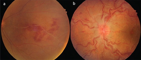
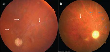
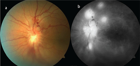
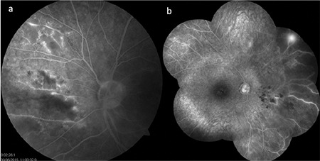
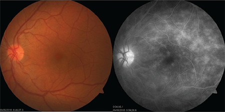
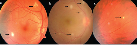
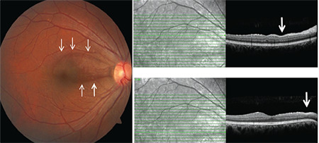
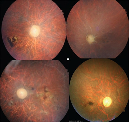
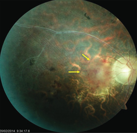
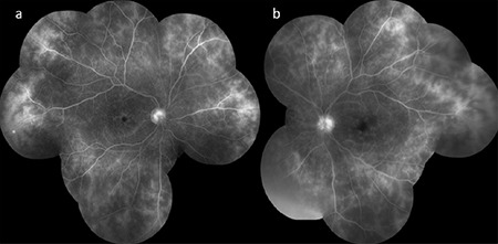
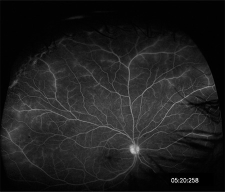
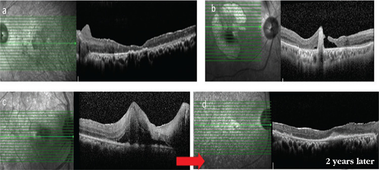
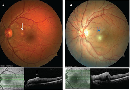
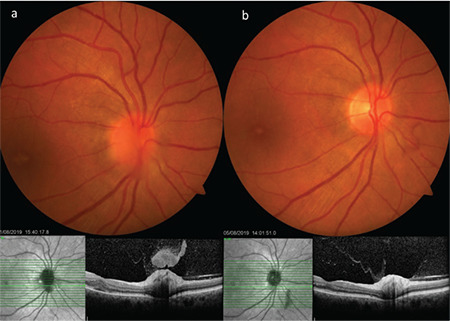
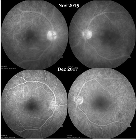
Behçet's disease is a chronic, multisystem inflammatory disorder characterized by relapsing inflammation. Although its etiopathogenesis has not yet been clarified, both the adaptive and innate immune systems, genetic predisposition, and environmental factors have all been implicated. It is more frequent and more severe in males in the third and fourth decades of life. The eye is the most frequently involved organ in the course of the disease. Ocular involvement (Behçet's uveitis) is characterized by bilateral recurrent non-granulomatous panuveitis and occlusive retinal vasculitis. Recurrent inflammatory episodes in the posterior segment may lead to permanent vision loss due to irreversible retinal damage and complications such as macular scarring, macular atrophy, and optic atrophy. Early and aggressive immunomodulatory treatment and the use of biologic agents when needed are crucial for preventing recurrences and improving visual prognosis.

Keywords: Behçet’s uveitis; biologics; imaging; prognosis; treatment.


Figures

Figure 1
Figure 1
Soft hypopyon (a) that moves freely (b) with head movements (red arrow) is seen in a patient with Behçet’s uveitis. Note that the eye is white despite hypopyon
Figure 2
Figure 2
Diffuse vitritis and vitreous haze are observed in two different Behçet’s uveitis patients. In the first patient (a), the optic disc is hyperemic and there is a small retinitis focus (white arrow) at the posterior pole. In the other patient (b), the vitreous haze is very dense and the optic disc (black arrow) and ghost vessels below (blue arrows) are barely discernible
Figure 3
Figure 3
In different patients (a,b,c,d), precipitates (vitreous pearls) are seen in the inferior periphery of the retina, indicating a regressing acute inflammatory episode
Figure 4
Figure 4
Different patients exhibit perivenous white sheathing (a, b, yellow arrows), hemorrhagic occlusive vasculitis at the posterior pole and associated hemorrhage in the papillomacular bundle (c, white arrow), and hemorrhage and exudates in the inferior macular region (d, white arrow)
Figure 5
Figure 5
Branch retinal vein occlusion (a) and central retinal vein occlusion (b) due to occlusive retinal vasculitis in two different Behçet’s patients
Figure 6
Figure 6
Gliotic sheathing (white arrows) of the retinal vessels and ghost vessels (blue arrows) are observed in two different Behçet’s patients (a, b)
Figure 7
Figure 7
Color fundus photographs (a) and fluorescein angiography images (b) of a Behçet’s patient who developed optic disc neovascularization. There is extensive vascular and capillary leakage
Figure 8
Figure 8
Fluorescein angiography images of two different Behçet’s uveitis patients with occlusive retinal vasculitis. The first (a) shows nasal optic disc ischemia and collateral formation in some areas; a composite image from the other patient (b) shows diffuse ischemia in the nasal periphery and retinal neovascularization
Figure 9
Figure 9
Fluorescein angiography of a Behçet’s patient with no clinically apparent retinal vasculitis on color fundus photograph shows optic disc staining, macular edema, and diffuse capillary and vascular leakage
Figure 10
Figure 10
Superficial retinal infiltrates (arrows) are observed in different patients (a, b, c). Even a single one (c) is regarded as posterior segment involvement
Figure 11
Figure 11
Color fundus photograph of a Behçet’s uveitis patient shows a wedgeshaped localized retinal nerve fiber layer loss (arrows) in the superior macula and the papillomacular bundle and thinning (arrow) on SD-OCT sections corresponding to the area of loss
Figure 12
Figure 12
The appearance of the fundus in different patients with end-stage disease. Optic atrophy, macular scarring, retinal atrophy, ghost vessels, and areas of retinal pigmentation can be seen
Figure 13
Figure 13
Active vasculitis in the papillomacular bundle (yellow arrows) is observed in an end-stage eye with retinal and macular atrophy, gliotic sheathing, pigmentation, and optic atrophy
Figure 14
Figure 14
Bilateral (a,b) optic disc staining, cystoid macular edema, vascular leakage, and fern-shaped capillary leakage are noted on fluorescein angiography in a Behçet’s patient
Figure 15
Figure 15
Fluorescein angiography with ultra-wide-field imaging shows vascular leakage in the superior and temporal periphery in addition to the optic disc and macular leakage. Shadowing caused by the lashes is present in the inferior and nasal regions
Figure 16
Figure 16
Macular atrophy in a patient with advanced Behçet’s uveitis (a), macular atrophy and hole in another patient (b), a patient who presented with active retinitis involving the macula and associated macular edema (c), and the same patient 2 years later, exhibiting disorganization and atrophy of the retinal layers and subfoveal fibrosis (d)
Figure 17
Figure 17
The superficial retinal infiltrates (a, white arrow; b, blue arrow) associated with Behçet’s uveitis led to focal retinal thickening and blurring and increased hyperreflectivity in the inner retinal layers in particular, while the contour of the retinal pigment epithelium was not disrupted
Figure 18
Figure 18
A patient with Behçet’s neuroretinitis exhibits optic disc infiltration accompanied by vitreous inflammation that looks like a hat over the disc on SDOCT, as well as subfoveal fluid and cystic edema (a). Four days after intravenous methylprednisolone therapy, substantial regression of the optic disc infiltration, overlying vitreous inflammation, and macular edema are observed (b)
Figure 19
Figure 19
A Behçet’s uveitis patient with bilateral optic disc staining, macular edema, and vascular and capillary leakage shows marked improvement approximately 2 years after interferon-alpha therapy

Similar articles

Cited by

References

    1. Tugal-Tutkun I. Behçet’s Uveitis. Middle East Afr J Ophthalmol. 2009;16:219–224. - PMC - PubMed
    1. Behçet H. Über rezidivierende aphthöse durch ein Virus verursachte Geschwüre am Mund, am Auge und an den Genitalien. Dermatol Wochenschr. 1937;105:1152–1157.
    1. Keino H, Okada AA. Behçet’s disease: global epidemiology of an Old Silk Road disease. Br J Ophthalmol. 2007;91:1573–1574. - PMC - PubMed
    1. Khairallah M, Accorinti M, Muccioli C, Kahloun R, Kempen JH. Epidemiology of Behçet disease. Ocul Immunol Inflamm. 2012;20:324–335. - PubMed
    1. Azizlerli G, Köse AA, Sarica R, Gül A, Tutkun IT, Kulaç M, Tunç R, Urgancioğlu M, Dişçi R. Prevalence of Behçet’s disease in Istanbul, Turkey. Int J Dermatol. 2003;42:803–806. - PubMed