Case Reports
. 2020 Jun 27;50(3):193-196.
doi: 10.4274/tjo.galenos.2020.92972.

Paracentral Acute Middle Maculopathy

Affiliations
Case Reports

Paracentral Acute Middle Maculopathy

Pelin Kıyat et al. Turk J Ophthalmol. .

Abstract

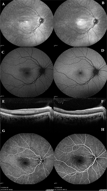
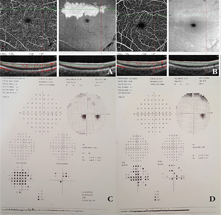
Paracentral acute middle maculopathy (PAMM) is a variant of acute macular neuroretinopathy which is characterized by a hyperreflective band-like lesion in the inner nuclear layer and outer plexiform layer on spectral domain optical coherence tomography (SD-OCT). The etiology is believed to involve vasopressor exposure or systemic microvascular diseases that cause retinal ischemia. SD-OCT is the main imaging method in the diagnosis or evaluation of progression of PAMM, whereas multimodal imaging is useful to support the diagnosis. Herein, we present a case of PAMM in a healthy young woman using multimodal imaging methods.

Keywords: Paracentral acute middle maculopathy; acute macular neuroretinopathy; optical coherence tomography.


Conflict of interest statement

Conflict of Interest: No conflict of interest was declared by the authors.

Figures

Figure 1
Figure 1
Fundus infrared, autofluorescence and SD-OCT and images at the time of diagnosis and at 4-month follow-up examination and FFA images at the time of diagnosis. In infrared images, hyporeflectance was detected in the superotemporal fovea lesion at the time of diagnosis (A) and was found to have regressed at 4-month follow-up (B). In fundus autofluorescence imaging, hypoautofluorescence was detected in the superotemporal fovea lesion at the time of diagnosis (C) and was found to have regressed at 4-month follow-up (D). SD-OCT performed in the right eye at time of diagnosis revealed a hyperreflective band lesion at the level of the inner nuclear and outer plexiform layer corresponding to the hypopigmented lesion observed in the superotemporal fovea (E). SD-OCT at 4-month follow-up demonstrated regression of the hyperreflective band including the inner nuclear and outer plexiform region in the superotemporal fovea, leaving retinal thinning and disorganization of the outer retinal layers in the lesion area (F). Early and late stage fundus fluorescein angiography images at the time of diagnosis (G-H). SD-OCT: Spectral domain optical coherence tomography
Figure 2
Figure 2
OCT angiography images and 30-2 visual field test at the time of diagnosis and at 4-month follow-up examination. OCT angiography at time of diagnosis demonstrated substantial capillary dropout in the deep capillary plexus (A). At 4-month follow-up, the capillary dropout was seen to have resolved, leaving disorganization in the deep capillary plexus (B). At diagnosis, 30-2 visual field test showed paracentral scotoma consistent with the hypopigmented lesion observed on fundus examination (C). On follow-up examination, the paracentral scotoma was found to have regressed (D). OCT: Optical coherence tomography

References

    1. Sarraf D, Rahimy E, Fawzi AA, Sohn E, Barbazetto I, Zacks DN, Mittra RA, Klancnik Jr JM, Mrejen S, Goldberg NR, Beardsley R, Sorenson JA, Freund KB. Paracentral acute middle maculopathy: A new variant of acute macular neuroretinopathy associated with retinal capillary ischemia. JAMA Ophthalmol. 2013;131:1275–1287. - PubMed
    1. Chu S, Nesper PL, Soetikno BT, Bakri SJ, Fawzi AA. Projection resolved oct angiography of microvascular changes in paracentral acute middlemaculopathy and acute macular neuroretinopathy. Invest Ophthalmol Vis Sci. 2018;59:2913–2922. - PMC - PubMed
    1. Shah D, Saurabh K, Roy R. Multimodal imaging in paracentral acute middle maculopathy. Indian J Ophthalmol. 2018;66:1186–1188. - PMC - PubMed
    1. El-Dairi M, Bhatti MT, Vaphiades MS. A shot of adrenaline. Surv Ophthalmol. 2009;54:618–624. - PubMed
    1. Yu S, Wang F, Pang CE, Yannuzzi LA, Freund KB. Multimodal imaging findings in retinal deep capillary ischemia. Retina. 2014;34:636–646. - PubMed

Publication types

LinkOut - more resources