Review
. 2020 Dec;11(1):132-148.
doi: 10.1080/19491034.2020.1782024.

CTCF as a boundary factor for cohesin-mediated loop extrusion: evidence for a multi-step mechanism

Affiliations
Review

CTCF as a boundary factor for cohesin-mediated loop extrusion: evidence for a multi-step mechanism

Anders S Hansen. Nucleus. 2020 Dec.

Abstract

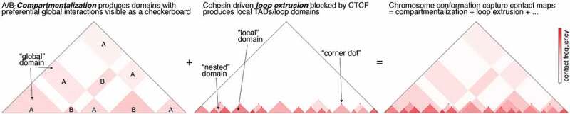
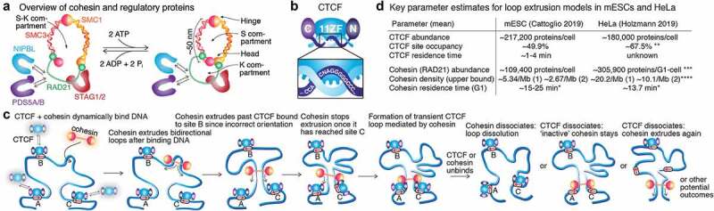
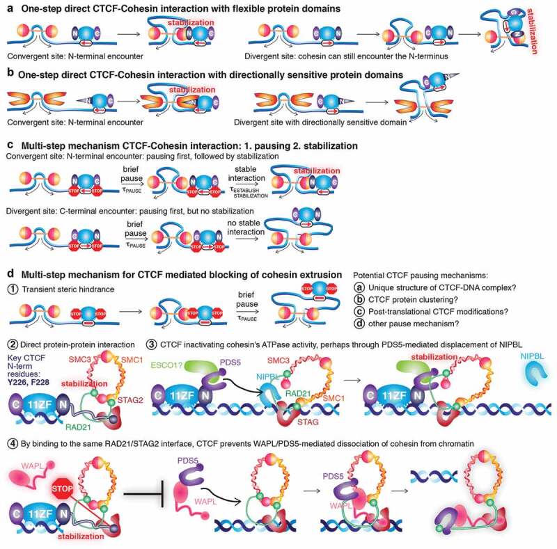
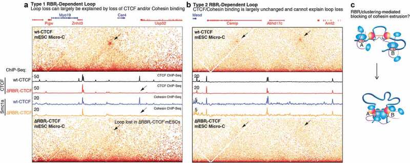
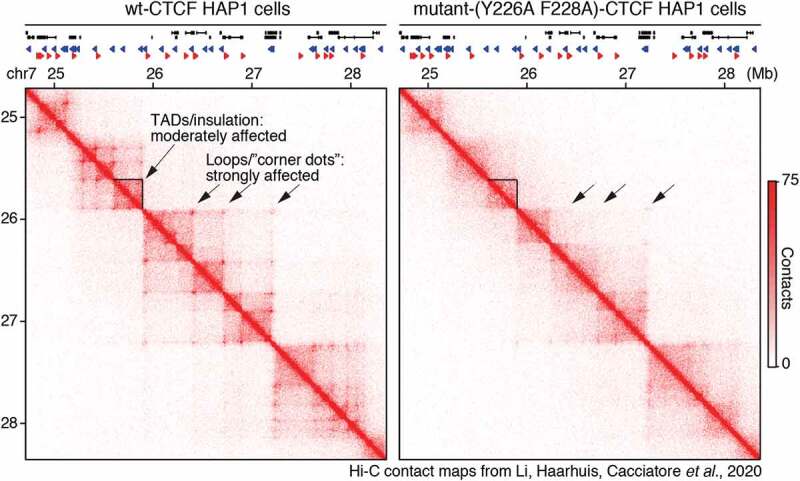
Mammalian genome structure is closely linked to function. At the scale of kilobases to megabases, CTCF and cohesin organize the genome into chromatin loops. Mechanistically, cohesin is proposed to extrude chromatin loops bidirectionally until it encounters occupied CTCF DNA-binding sites. Curiously, loops form predominantly between CTCF binding sites in a convergent orientation. How CTCF interacts with and blocks cohesin extrusion in an orientation-specific manner has remained a mechanistic mystery. Here, we review recent papers that have shed light on these processes and suggest a multi-step interaction between CTCF and cohesin. This interaction may first involve a pausing step, where CTCF halts cohesin extrusion, followed by a stabilization step of the CTCF-cohesin complex, resulting in a chromatin loop. Finally, we discuss our own recent studies on an internal RNA-Binding Region (RBRi) in CTCF to elucidate its role in regulating CTCF clustering, target search mechanisms and chromatin loop formation and future challenges.

Keywords: Binding Polarity; CTCF; Cohesin; Convergent Rule; Loop Extrusion; NIPBL; PDS5; RNA-Binding Region; TADs.


Conflict of interest statement

I declare that no competing financial interests exist.

Figures

Figure 1.
Figure 1.
A simplified illustration of how contact map features are shaped by A/B-compartmentalization and loop extrusion. Highly simplified sketches of hypothetical contact maps produced by chromosome conformation capture methods such as Hi-C. Left: Hypothetical contact map produced by A/B-compartmentalization. Compartmentalization generates both local and global domains. Middle: Hypothetical contact map produced by loop extrusion. Loop extrusion generates strictly local maps demarcated by strong convergent CTCF binding sites, and sometimes forms nested domains. Right: Real contact maps are affected by both A/B-compartmentalization and CTCF/cohesin-mediated loop extrusion – as well as a number of other processes especially at the fine-scale [129,130] – and are therefore the sum of all of these processes. This makes interpreting and classifying ‘domains’ in Hi-C contact maps highly challenging [9,30].
Figure 2.
Figure 2.
Overview of cohesin, CTCF, and loop extrusion. (a) Overview of mammalian cohesin and some of its regulatory proteins. (b) Overview of CTCF with N-terminal, 11 Zinc Fingers, and C-terminal domains. (c) Simplified sketch of cohesin-mediated loop extrusion and the convergent CTCF rule. (d) Summary of key parameters constraining loop extrusion models in mouse embryonic stem cells (mESCs) [65] and human HeLa cells [83], with mESC residence times taken from [70]. * These are cohesin G1 residence times (both STAG1 and STAG2), but after these studies were published it was found that STAG2-cohesin binds DNA substantially more dynamically than STAG1-cohesin [38], suggesting that putative loop extruding G1 cohesins have at least two residence times. ** Estimated from [83] (~180,000 and ~120,000 CTCF proteins and sites per HeLa cell) with added assumption that 45% of CTCF proteins are bound to cognate sites (~45%, i.e. mean of mESC and U2OS in [70]). *** 305,900 is the mean of the LC-MS and FCS estimates reported in [83]. **** Cohesin density is estimated from ~159,437 dynamically bound (~13.7 min residence time) cohesin proteins (SCC1-mEGFP) in G1 and the reported HeLa genome sizes 7.9 Gb, both taken from [83]. It is important to note that these are genomic averages: e.g. CTCF residence time is for an average site (some sites will have slower and faster binding), cohesin density may not be uniform throughout the genome, and since the two in vitro cohesin loop extrusion papers disagreed on whether cohesin is monomeric [23] or dimeric [21], densities for both monomeric [1] and dimeric [2] are shown.
Figure 3.
Figure 3.
One-step vs. Multi-step CTCF-cohesin interaction mechanisms. (a) One-step CTCF-cohesin mechanism. If 1-step mechanism, it is not clear why cohesin could not extrude past the C-terminal domain of CTCF to interact with the N-terminal domain on the other side. (b) One-step CTCF-cohesin mechanism with directionally sensitive domains. For a one-step mechanism to work, the N-terminal CTCF domain and cohesin would both have to exhibit a directional sensitivity as illustrated. (c) Multi-step CTCF-cohesin mechanism. For an N-terminal encounter, pausing is eventually followed by stabilization. For a C-terminal encounter, pausing is not followed by stabilization, so cohesin eventually extrudes past or dissociates. (d) Instead of a one-step mechanism, a multi-step mechanism would involve transient pausing of cohesin next to CTCF (1), followed by stabilization of cohesin only from the N-terminal side of CTCF, through either direct protein-protein interaction (2), CTCF ‘turning OFF’ the cohesin motor (ATPase) perhaps mediated via PDS5A/B and/or ESCO1 (3), or through CTCF preventing WAPL-mediated release of cohesin from chromatin by CTCF binding to the same RAD21/STAG2 interface as WAPL does (4). It is important to note both that these mechanisms are not mutually exclusive, and that many other mechanisms could contribute.
Figure 4.
Figure 4.
Distinct classes of chromatin loops. (a-b) Micro-C maps and CTCF and Cohesin (Smc1a) ChIP-Seq shown for wt-CTCF mESCs and ΔRBRi-CTCF mESCs, illustrating Type 1 RBRi-dependent loops that can be explained by loss of CTCF/cohesin binding (a) and Type 2 RBRi-dependent loops that cannot be explained by loss of CTCF/cohesin binding (b). (c) Sketch of a role for CTCF clustering in blocking cohesin extrusion. Figures 4a-c are partially reproduced and edited from [36] with permission.
Figure 5.
Figure 5.
CTCF-mediated loops can be disrupted with only modest effects on TADs/insulation. (a-b) Hi-C contact matrices at 10 kb resolution of the HOXA locus in wt-CTCF and Y226A/F228A-CTCF HAP1 cells from [40].

Similar articles

Cited by

References

    1. Cremer T, Cremer C.. Chromosome territories, nuclear architecture and gene regulation in mammalian cells. Nat Rev Genet. 2001;2(4):292–301. - PubMed
    1. Luger K, Mäder AW, Richmond RK, et al. Crystal structure of the nucleosome core particle at 2.8 Å resolution. Nature. 1997;389(6648):251–260. - PubMed
    1. Nuebler J, Fudenberg G, Imakaev M, et al. Chromatin organization by an interplay of loop extrusion and compartmental segregation. Proceed Nat Acad Sci. 2018;115(29):E6697–E6706. - PMC - PubMed
    1. Lieberman-Aiden E, van Berkum NL, Williams L, et al. Science. Comprehensive mapping of long-range interactions reveals folding principles of the human genome. Science. 2009;326(5950):289–293. - PMC - PubMed
    1. Hildebrand EM, Dekker J. Mechanisms and functions of chromosome compartmentalization. Trends Biochem Sci. 2020;45(5):385–396. - PMC - PubMed

Publication types