. 2020 Jul 6;8(1):99.
doi: 10.1186/s40478-020-00972-z.

Disturbed balance in the expression of MMP9 and TIMP3 in cerebral amyloid angiopathy-related intracerebral haemorrhage

Affiliations

Disturbed balance in the expression of MMP9 and TIMP3 in cerebral amyloid angiopathy-related intracerebral haemorrhage

Lieke Jäkel et al. Acta Neuropathol Commun. .

Abstract

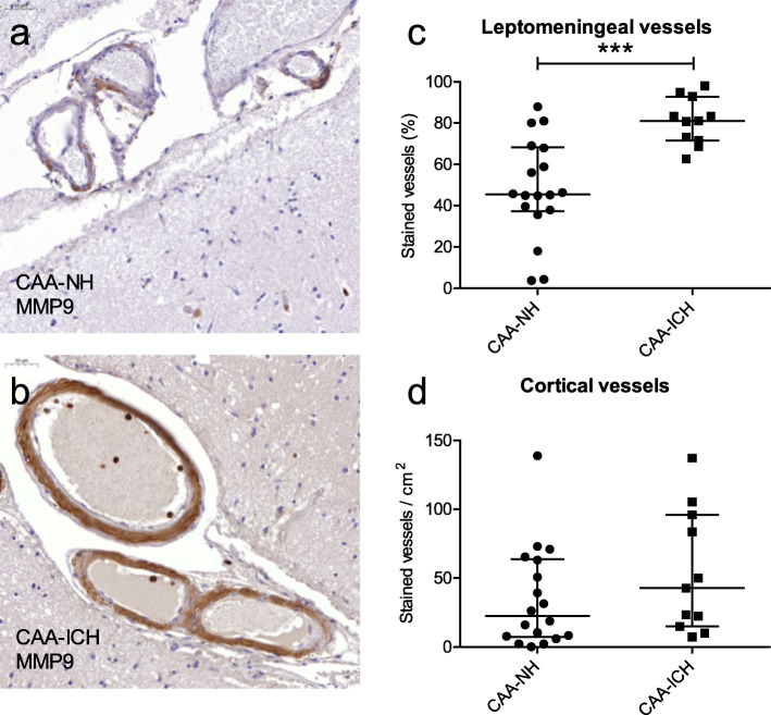
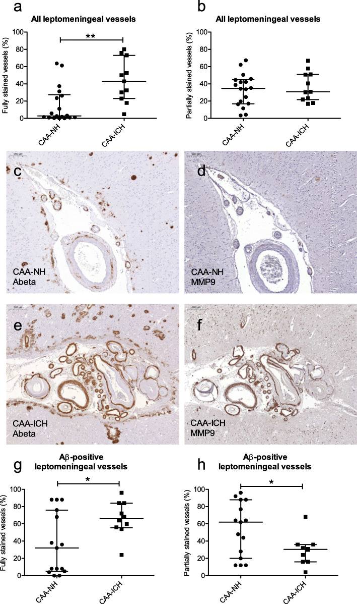
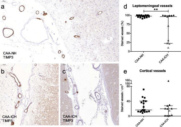
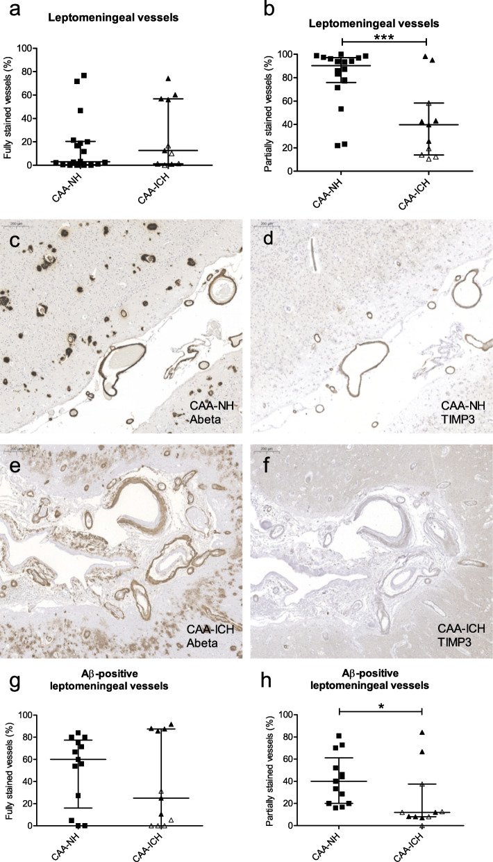
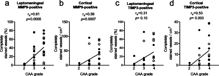
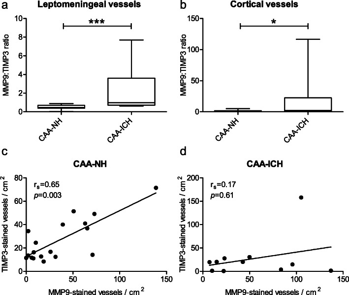
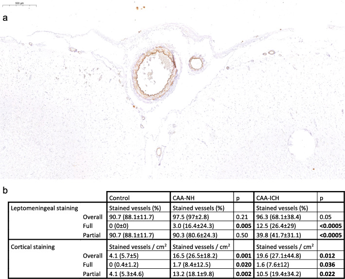
Cerebral amyloid angiopathy (CAA) is characterized by the deposition of the amyloid β (Aβ) protein in the cerebral vasculature and poses a major risk factor for the development of intracerebral haemorrhages (ICH). However, only a minority of patients with CAA develops ICH (CAA-ICH), and to date it is unclear which mechanisms determine why some patients with CAA are more susceptible to haemorrhage than others. We hypothesized that an imbalance between matrix metalloproteinases (MMPs) and their inhibitors (TIMPs) contributes to vessel wall weakening. MMP9 plays a role in the degradation of various components of the extracellular matrix as well as of Aβ and increased MMP9 expression has been previously associated with CAA. TIMP3 is an inhibitor of MMP9 and increased TIMP3 expression in cerebral vessels has also been associated with CAA. In this study, we investigated the expression of MMP9 and TIMP3 in occipital brain tissue of CAA-ICH cases (n = 11) by immunohistochemistry and compared this to the expression in brain tissue of CAA cases without ICH (CAA-non-haemorrhagic, CAA-NH, n = 18). We showed that MMP9 expression is increased in CAA-ICH cases compared to CAA-NH cases. Furthermore, we showed that TIMP3 expression is increased in CAA cases compared to controls without CAA, and that TIMP3 expression is reduced in a subset of CAA-ICH cases compared to CAA-NH cases. In conclusion, in patients with CAA, a disbalance in cerebrovascular MMP9 and TIMP3 expression is associated with CAA-related ICH.

Keywords: Alzheimer’s disease; Amyloid β protein; Cerebral amyloid angiopathy; Intracerebral haemorrhage; Matrix metalloproteinase 9; Tissue inhibitor of metalloproteinases 3.


Conflict of interest statement

The authors declare that they have no competing interests.

Figures

Fig. 1
Fig. 1
Cerebrovascular MMP9 expression in CAA-NH and CAA-ICH cases. Representative examples of MMP9 staining in a CAA-NH case (a), and a CAA-ICH case (b), representing the median values as shown in c. Leptomeningeal vessels are stained in both cases: in CAA-NH many vessels are only partially stained, whereas in CAA-ICH cases many vessels are fully stained. Overall, CAA-ICH cases had a significantly higher percentage of MMP9-stained leptomeningeal vessels (either fully or partially stained) compared to CAA-NH cases (c). The numbers of MMP9-stained cortical vessels did not differ between groups (d). Plots indicate median values with interquartile range. CAA-NH = CAA-non haemorrhagic, CAA-ICH = CAA-related ICH. ***p ≤ 0.001
Fig. 2
Fig. 2
Quantification of MMP9-stained vessels in CAA-NH and CAA-ICH cases, classified according to staining grade. The percentage of leptomeningeal vessels with full (a), but not partial (b) MMP9 staining was higher in CAA-ICH cases compared to CAA-NH cases. Serial sections of a representative CAA-NH (c, d) and a representative CAA-ICH (e, f) case stained for Aβ (c, e) and MMP9 (d, f). Scale bar = 200 μm. Compared to CAA-NH cases, CAA-ICH cases had more fully Aβ-stained leptomeningeal vessels that were also fully stained for MMP9 (g), but fewer Aβ-stained vessels that had partial MMP9 staining (h). Plots indicate median values with interquartile range. CAA-NH = CAA-non haemorrhagic, CAA-ICH = CAA-related ICH. *p ≤ 0.05; **p ≤ 0.01
Fig. 3
Fig. 3
Cerebrovascular TIMP3 expression in CAA-NH and CAA-ICH cases. Representative example of TIMP3 staining in a CAA-NH case (a), a CAA-ICH case with high TIMP3 expression (b), and a CAA-ICH case with many vessels negative for TIMP3 and only few TIMP3- positive vessels (c). Scale bar = 100 μm (a). The percentage of TIMP3-stained (full or partial) leptomeningeal vessels was lower in CAA-ICH cases compared to CAA-NH cases, an effect driven by a subgroup of CAA-ICH cases with a substantially lower percentage of TIMP3-stained vessels (open triangles) (b). The numbers of cortical vessels stained per cm2 did not differ between CAA-ICH cases and CAA-NH cases, although the cases with low TIMP3 expression in leptomeningeal vessels had also low expression in cortical vessels (open triangles) (c). Plots indicate median values with interquartile range. CAA-NH = CAA-non haemorrhagic, CAA-ICH = CAA-related ICH. **p ≤ 0.01
Fig. 4
Fig. 4
Quantification of TIMP3-stained vessels in CAA-NH and CAA-ICH cases, classified according to staining grade. The percentage of leptomeningeal vessels with full TIMP3 staining did not differ between groups (a), whereas the percentage with partial staining was significantly lower in the CAA-ICH group (b). Serial sections stained for Aβ (c, e) and TIMP3 (d, f) in a CAA-NH case (c, d) and a CAA-ICH case with low levels of TIMP3 expression (e, f) showed that these proteins colocalize. Scale bar = 200 μm. Assessment of only fully Aβ-stained leptomeningeal vessels showed no difference in the percentage of vessels that had full TIMP3 staining (g), but a lower percentage of partially stained vessels in the CAA-ICH group (h). The observed differences were predominantly driven by the subgroup of CAA-ICH cases with low TIMP3 expression (open triangles). Plots indicate median values with interquartile range. CAA-NH = CAA-non haemorrhagic, CAA-ICH = CAA-related ICH, *p ≤ 0.05; ***p ≤ 0.001
Fig. 5
Fig. 5
The expression of MMP9 and TIMP3-stained vessels correlated with CAA grade. The percentage of fully MMP9-stained leptomeningeal (a) and number of fully MMP9-stained cortical (b) vessels showed a positive correlation with CAA grade. The percentage of fully TIMP3-stained leptomeningeal vessels did not significantly correlate to CAA grade (c), whereas the numbers of fully TIMP3-stained cortical (d) vessels showed a positive correlation with CAA grade. Solid circles = CAA-NH cases; open circles = CAA-ICH cases; rs = Spearman r
Fig. 6
Fig. 6
Disturbed balance of MMP9 and TIMP3 in vessels of CAA-ICH cases. The ratio of MMP9-stained (partially or fully) to TIMP3-stained (partially or fully) leptomeningeal vessels (a) and  cortical vessels (b) was higher in CAA-ICH cases compared to CAA-NH casesIn CAA-NH cases, there was a positive correlation between the numbers of cortical vessels stained (partially or fully) for MMP9 and the numbers of cortical vessels stained (partially or fully) for TIMP3 (c). Such a correlation was not seen in CAA-ICH cases (d). Box plots show median values with the 25th and 75th percentile as boundaries and whiskers indicating minimum and maximum values. CAA-NH = CAA-non haemorrhagic, CAA-ICH = CAA-related ICH, *p ≤ 0.05; ***p ≤ 0.001, rs = Spearman r
Fig. 7
Fig. 7
TIMP3 staining in control cases. Representative example of TIMP3 staining in a control case (a). Table shows median (mean ± sd) values of the percentages (in case of leptomeningeal) or numbers per cm2 (in case of cortical) vessels (b). The difference in expression between control cases and CAA-NH or CAA-ICH cases was assessed by linear regression with age and sex as covariates. Indicated in bold are p-values < 0.05. CAA-NH = CAA-non haemorrhagic, CAA-ICH = CAA-related ICH

References

    1. Attems J. Sporadic cerebral amyloid angiopathy: pathology, clinical implications, and possible pathomechanisms. Acta Neuropathol. 2005;110:345–359. doi: 10.1007/s00401-005-1074-9. - DOI - PubMed
    1. Attems J, Jellinger KA, Lintner F. Alzheimer's disease pathology influences severity and topographical distribution of cerebral amyloid angiopathy. Acta Neuropathol. 2005;110:222–231. doi: 10.1007/s00401-005-1064-y. - DOI - PubMed
    1. Attems J, Lauda F, Jellinger KA. Unexpectedly low prevalence of intracerebral hemorrhages in sporadic cerebral amyloid angiopathy: an autopsy study. J Neurol. 2008;255:70–76. doi: 10.1007/s00415-008-0674-4. - DOI - PubMed
    1. Backstrom JR, Lim GP, Cullen MJ, Tokes ZA. Matrix metalloproteinase-9 (MMP-9) is synthesized in neurons of the human hippocampus and is capable of degrading the amyloid-beta peptide (1-40) J Neurosci. 1996;16:7910–7919. doi: 10.1523/JNEUROSCI.16-24-07910.1996. - DOI - PMC - PubMed
    1. Banerjee G, Carare R, Cordonnier C, Greenberg SM, Schneider JA, Smith EE, et al. The increasing impact of cerebral amyloid angiopathy: essential new insights for clinical practice. J Neurol Neurosurg Psychiatry. 2017;88:982–994. doi: 10.1136/jnnp-2016-314697. - DOI - PMC - PubMed

Publication types

Substances

LinkOut - more resources