

Validation of the British Society of Thoracic Imaging guidelines for COVID-19 chest radiograph reporting
- PMID: 32631626
- PMCID: PMC7298474
- DOI: 10.1016/j.crad.2020.06.005
Validation of the British Society of Thoracic Imaging guidelines for COVID-19 chest radiograph reporting
Abstract
Aim: To validate the British Society of Thoracic Imaging issued guidelines for the categorisation of chest radiographs for coronavirus disease 2019 (COVID-19) reporting regarding reproducibility amongst radiologists and diagnostic performance.
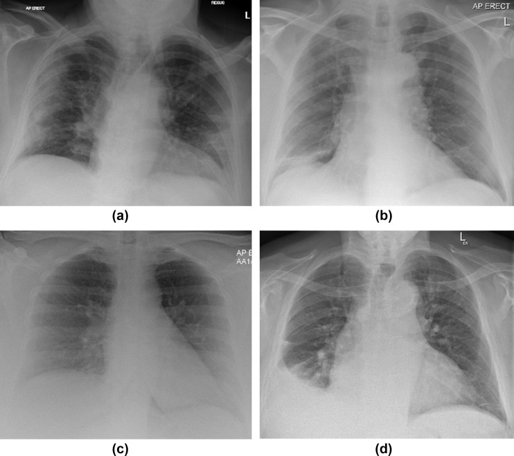
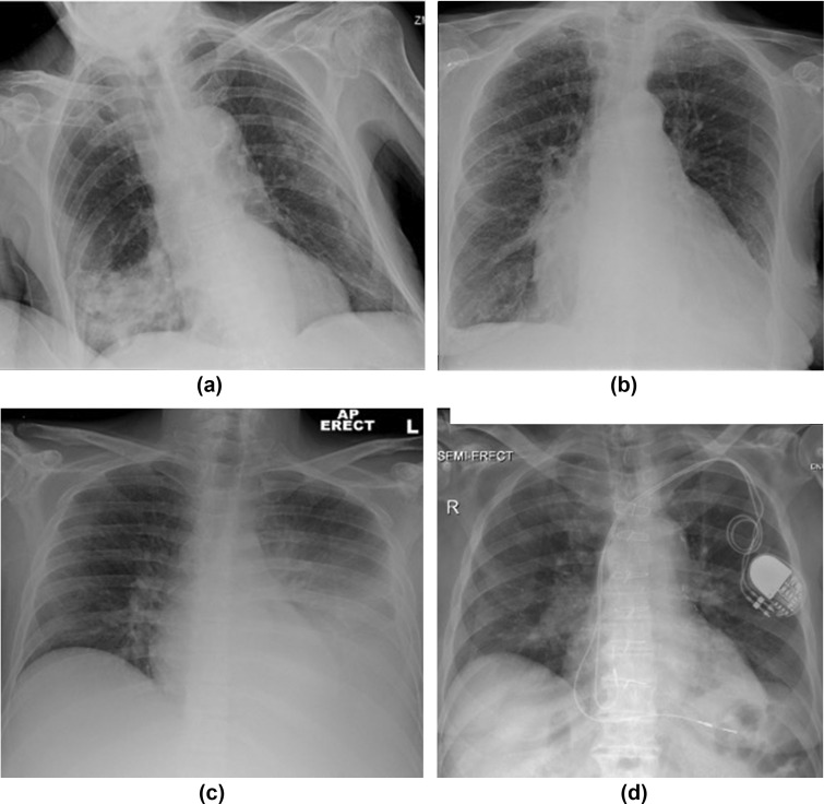
Materials and methods: Chest radiographs from 50 patients with COVID-19, and 50 control patients with symptoms consistent with COVID-19 from prior to the emergence of the novel coronavirus were assessed by seven consultant radiologists with regards to the British Society of Thoracic Imaging guidelines.
Results: The findings show excellent specificity (100%) and moderate sensitivity (44%) for guideline-defined Classic/Probable COVID-19, and substantial interobserver agreement (Fleiss' k=0.61). Fair agreement was observed for the "Indeterminate for COVID-19" (k=0.23), and "Non-COVID-19" (k=0.37) categories; furthermore, the sensitivity (0.26 and 0.14 respectively) and specificity (0.76, 0.80) of these categories for COVID-19 were not significantly different (McNemar's test p=0.18 and p=0.67).
Conclusion: An amalgamation of the categories of "Indeterminate for COVID-19" and "Non-COVID-19" into a single "not classic of COVID-19" classification would improve interobserver agreement, encompass patients with a similar probability of COVID-19, and remove the possibility of labelling patients with COVID-19 as "Non-COVID-19", which is the presenting radiographic appearance in a significant minority (14%) of patients.
Copyright © 2020. Published by Elsevier Ltd.
Conflict of interest statement
Conflicts of interest Dr Hare is on the committee of the British Society of Thoracic Imaging.
Figures
Similar articles
-
Canadian Society of Thoracic Radiology/Canadian Association of Radiologists Consensus Statement Regarding Chest Imaging in Suspected and Confirmed COVID-19.Can Assoc Radiol J. 2020 Nov;71(4):470-481. doi: 10.1177/0846537120924606. Epub 2020 May 8. Can Assoc Radiol J. 2020. PMID: 32380844
-
Chest CT and Coronavirus Disease (COVID-19): A Critical Review of the Literature to Date.AJR Am J Roentgenol. 2020 Oct;215(4):839-842. doi: 10.2214/AJR.20.23202. Epub 2020 Apr 16. AJR Am J Roentgenol. 2020. PMID: 32298149 Review.
-
Thoracic imaging tests for the diagnosis of COVID-19.Cochrane Database Syst Rev. 2020 Sep 30;9:CD013639. doi: 10.1002/14651858.CD013639.pub2. Cochrane Database Syst Rev. 2020. Update in: Cochrane Database Syst Rev. 2020 Nov 26;11:CD013639. doi: 10.1002/14651858.CD013639.pub3. PMID: 32997361 Updated.
-
Chest x-ray in the COVID-19 pandemic: Radiologists' real-world reader performance.Eur J Radiol. 2020 Nov;132:109272. doi: 10.1016/j.ejrad.2020.109272. Epub 2020 Sep 10. Eur J Radiol. 2020. PMID: 32971326 Free PMC article.
-
Thoracic imaging of coronavirus disease 2019 (COVID-19) in children: a series of 91 cases.Pediatr Radiol. 2020 Sep;50(10):1354-1368. doi: 10.1007/s00247-020-04747-5. Epub 2020 Aug 4. Pediatr Radiol. 2020. PMID: 32749530 Free PMC article.
Cited by
-
Sensitivity of SARS-CoV-2 RNA polymerase chain reaction using a clinical and radiological reference standard.J Infect. 2021 Jun;82(6):260-268. doi: 10.1016/j.jinf.2021.04.012. Epub 2021 Apr 20. J Infect. 2021. PMID: 33892014 Free PMC article.
-
Preoperative COVID-19 CT screening in renal transplant recipients.Clin Radiol. 2020 Nov;75(11):868-870. doi: 10.1016/j.crad.2020.08.004. Epub 2020 Aug 14. Clin Radiol. 2020. PMID: 32868090 Free PMC article. No abstract available.
-
Review on Diagnosis of COVID-19 from Chest CT Images Using Artificial Intelligence.Comput Math Methods Med. 2020 Sep 26;2020:9756518. doi: 10.1155/2020/9756518. eCollection 2020. Comput Math Methods Med. 2020. PMID: 33014121 Free PMC article. Review.
-
Chest radiography findings of COVID-19 pneumonia: a specific pattern for a confident differential diagnosis.Acta Radiol. 2022 Dec;63(12):1619-1626. doi: 10.1177/02841851211055163. Epub 2021 Nov 13. Acta Radiol. 2022. PMID: 34779269 Free PMC article.
-
Assessment of COVID-19 RT-PCR Positive Symptomatic Patients With Clinical, Hematological, and Radiological Parameters Among Three Groups: A Comparative Study.Cureus. 2023 May 30;15(5):e39681. doi: 10.7759/cureus.39681. eCollection 2023 May. Cureus. 2023. PMID: 37398817 Free PMC article.
References
-
- NHS England Specialty guides for patient management during the coronavirus pandemic Clinical guide for the management of Radiology patients during the coronavirus pandemic. https://www.england.nhs.uk/coronavirus/wp-content/uploads/sites/52/2020/... Available at:
