Serial Molecular Profiling of Low-grade Prostate Cancer to Assess Tumor Upgrading: A Longitudinal Cohort Study
- PMID: 32631746
- PMCID: PMC7779657
- DOI: 10.1016/j.eururo.2020.06.041
Serial Molecular Profiling of Low-grade Prostate Cancer to Assess Tumor Upgrading: A Longitudinal Cohort Study
Abstract
Background: The potential for low-grade (grade group 1 [GG1]) prostate cancer (PCa) to progress to high-grade disease remains unclear.
Objective: To interrogate the molecular and biological features of low-grade PCa serially over time.
Design, setting, and participants: Nested longitudinal cohort study in an academic active surveillance (AS) program. Men were on AS for GG1 PCa from 2012 to 2017.
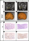
Intervention: Electronic tracking and resampling of PCa using magnetic resonance imaging/ultrasound fusion biopsy.
Outcome measurements and statistical analysis: ERG immunohistochemistry (IHC) and targeted DNA/RNA next-generation sequencing were performed on initial and repeat biopsies. Tumor clonality was assessed. Molecular data were compared between men who upgraded and those who did not upgrade to GG ≥ 2 cancer.
Results and limitations: Sixty-six men with median age 64 yr (interquartile range [IQR], 59-69) and prostate-specific antigen 4.9 ng/mL (IQR, 3.3-6.4) underwent repeat sampling of a tracked tumor focus (median interval, 11 mo; IQR, 6-13). IHC-based ERG fusion status was concordant at initial and repeat biopsies in 63 men (95% vs expected 50%, p < 0.001), and RNAseq-based fusion and isoform expression were concordant in nine of 13 (69%) ERG+ patients, supporting focal resampling. Among 15 men who upgraded with complete data at both time points, integrated DNA/RNAseq analysis provided evidence of shared clonality in at least five cases. Such cases could reflect initial undersampling, but also support the possibility of clonal temporal progression of low-grade cancer. Our assessment was limited by sample size and use of targeted sequencing.
Conclusions: Repeat molecular assessment of low-grade tumors suggests that clonal progression could be one mechanism of upgrading. These data underscore the importance of serial tumor assessment in men pursuing AS of low-grade PCa.
Patient summary: We performed targeted rebiopsy and molecular testing of low-grade tumors on active surveillance. Our findings highlight the importance of periodic biopsy as a component of monitoring for cancer upgrading during surveillance.
Keywords: Cancer progression; Gene fusions; Immunohistochemistry; Low-grade cancer; Next-generation sequencing; Prostate cancer; Tumor clonality.
Copyright © 2020 European Association of Urology. Published by Elsevier B.V. All rights reserved.
Figures


Comment in
-
Re: Simpa S. Salami, Jeffrey J. Tosoian, Srinivas Nallandhighal, et al. Serial Molecular Profiling of Low-grade Prostate Cancer to Assess Tumor Upgrading: A Longitudinal Cohort Study. Eur Urol. In press. https://doi.org/10.1016/j.eururo.2020.06.041.Eur Urol. 2021 Mar;79(3):e98-e99. doi: 10.1016/j.eururo.2020.12.001. Epub 2020 Dec 16. Eur Urol. 2021. PMID: 33341284 No abstract available.
-
Evidence for Focal Grade Group Progression in Low-risk Prostate Cancer.Eur Urol. 2021 Apr;79(4):466-467. doi: 10.1016/j.eururo.2020.11.022. Epub 2020 Dec 24. Eur Urol. 2021. PMID: 33357993 No abstract available.
References
-
- Klotz L, Vesprini D, Sethukavalan P, et al. Long-term follow-up of a large active surveillance cohort of patients with prostate cancer. J Clin Oncol 2015;33:272–7. - PubMed
-
- Hamdy FC, Donovan JL, Lane JA, et al. 10-Year outcomes after monitoring, surgery, or radiotherapy for localized prostate cancer. N Engl J Med 2016;375:1415–24. - PubMed
-
- Tosoian JJ, Mamawala M, Epstein JI, et al. Active surveillance of grade group 1 prostate cancer: long-term outcomes from a large prospective cohort. Eur Urol 2020;77:675–82. - PubMed
-
- Mohler J, Srinivas S, Antonarakis ES. NCCN clinical practice guidelines in oncology: prostate cancer. 2020. https://www.nccn.org/professionals/physician_gls/pdf/prostate.pdf
-
- Löppenberg B, Friedlander DF, Krasnova A, et al. Variation in the use of active surveillance for low-risk prostate cancer. Cancer 2018;124:55–64. - PubMed
Publication types
MeSH terms
Grants and funding
LinkOut - more resources
Full Text Sources
Other Literature Sources
Medical
Research Materials

