. 2020 Oct;26(10):1464-1480.
doi: 10.1261/rna.075655.120. Epub 2020 Jul 6.

Origin, conservation, and loss of alternative splicing events that diversify the proteome in Saccharomycotina budding yeasts

Affiliations

Origin, conservation, and loss of alternative splicing events that diversify the proteome in Saccharomycotina budding yeasts

Jennifer E Hurtig et al. RNA. 2020 Oct.

Abstract

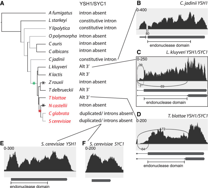
Many eukaryotes use RNA processing, including alternative splicing, to express multiple gene products from the same gene. The budding yeast Saccharomyces cerevisiae has been successfully used to study the mechanism of splicing and the splicing machinery, but alternative splicing in yeast is relatively rare and has not been extensively studied. Alternative splicing of SKI7/HBS1 is widely conserved, but yeast and a few other eukaryotes have replaced this one alternatively spliced gene with a pair of duplicated, unspliced genes as part of a whole genome doubling (WGD). We show that other examples of alternative splicing known to have functional consequences are widely conserved within Saccharomycotina. A common mechanism by which alternative splicing has disappeared is by replacement of an alternatively spliced gene with duplicate unspliced genes. This loss of alternative splicing does not always take place soon after duplication, but can take place after sufficient time has elapsed for speciation. Saccharomycetaceae that diverged before WGD use alternative splicing more frequently than S. cerevisiae, suggesting that WGD is a major reason for infrequent alternative splicing in yeast. We anticipate that WGDs in other lineages may have had the same effect. Having observed that two functionally distinct splice-isoforms are often replaced by duplicated genes allowed us to reverse the reasoning. We thereby identify several splice isoforms that are likely to produce two functionally distinct proteins because we find them replaced by duplicated genes in related species. We also identify some alternative splicing events that are not conserved in closely related species and unlikely to produce functionally distinct proteins.

Keywords: alternative splicing; evolution; subfunctionalization; yeast.


Figures

FIGURE 1.
FIGURE 1.
Alternative splicing of PTC7 orthologs in the Saccharomycotina. (A) Splicing pattern is shown for each of the species analyzed, along with the species phylogeny. Key branches are indicated: the branch leading to the subphylum Saccharomycotina, the branch leading to the family Saccharomycetacea, and the branch leading to the WGD clade. Red species names highlight the WGD clade. Green asterisk indicates the most likely origin of PTC7 intron retention. Red asterisk indicates the most likely replacement of one alternatively spliced gene with duplicate genes. (BG) Sashimi plots of a representative RNA-seq data set for the indicated species. Each plot indicates in the top left corner the read coverage scale on the y-axis. Arches indicate introns that were spliced out, and the numbers along the arches indicate the number of exon junction reads for that splicing pattern. Below each sashimi plot is the likely RNA structure with boxes indicating coding exons and lines indicating intron. TM indicates a transmembrane helix predicted to be encoded by the retained intron. (B) C. albicans diverged before the likely origin of PTC7 alternative splicing and no alternative splicing was detected. (C) Intron retention in the PTC7 ortholog from C. jadinii. (D) Intron retention in the PTC7 ortholog from T. delbrueckii. (E,F) T. blattae contains two PTC7 orthologs, with one lacking an intron and the other containing a constitutive intron. (G) Intron retention in PTC7 of S. cerevisiae. The box around this panel indicates this panel confirms the previously described alternative splicing.
FIGURE 2.
FIGURE 2.
Alternative splicing of MDH orthologs in the Saccharomycotina and A. fumigatus. (A) Splicing pattern is shown for each of the species analyzed, along with the species phylogeny as in Figure 1A. In addition, the black asterisks indicate a change of splicing pattern. Specifically, the ancestor likely used both intron retention and alternative 3′ splice sites. (*) Loss of alternative 3′ splice sites in L. starkeyi, loss of intron retention in Y. lipolytica, and the conversion of the peroxisomal gene into a pseudogene in O. polymorpha. (B) Intron retention and alternative 3′ splice sites in the A. fumigatus MDH gene. The light gray box indicates a reading frame that is different from the dark gray boxes. PTS indicates a peroxisomal targeting signal. (C) Intron retention (likely coupled with an intronic polyadenylation site) in the L. starkeyi MDH gene. (D) Alternative 3′ splice sites in the Y. lipolytica MDH gene. (E,F) Duplicated MDH genes in S. cerevisiae lack introns. In panels BD, only the 3′ end of the gene is shown.
FIGURE 3.
FIGURE 3.
Alternative splicing of YSH1/SYC1 orthologs in the Saccharomycotina. (A) Splicing pattern is shown for each of the species analyzed, along with the species phylogeny as in Figure 1A. Black asterisks indicate that Z. rouxii and N. castellii appear to have lost Syc1. (B) C. jadinii diverged before the likely origin of YSH1/SYC1 alternative splicing and its ortholog contains a constitutive intron. (C) Alternative 3′ splice sites in the YSH1/SYC1 ortholog of L. kluyveri, as previously reported. (D) Alternative 3′ splice sites in the YSH1/SYC1 ortholog of T. blattae. (E,F) Duplicated YSH1 and SYC1 genes in S. cerevisiae lack introns.
FIGURE 4.
FIGURE 4.
Alternative splicing of NUP116/NUP100 orthologs in the Saccharomycotina. (A) Splicing pattern is shown for each of the species analyzed, along with the species phylogeny as in Figure 1A. (B) C. jadinii diverged before the likely origin of NUP116/NUP100 alternative splicing and its ortholog lacks an intron. The C. jadinii ortholog contains a sequence that is homologous to the sequence in Nup116 that binds to Gle2. (C) Intron retention in the NUP116/NUP100 ortholog of L. kluyveri. (D) Intron retention in the NUP116/NUP100 ortholog of T. delbrueckii. (E,F) Duplicated NUP116 and NUP100 genes in S. cerevisiae lack introns and only Nup116 contains the Gle2 binding site.
FIGURE 5.
FIGURE 5.
Alternative splicing of GND orthologs in the Saccharomycotina and A. fumigatus. (A) Splicing pattern is shown for each of the species analyzed, along with the species phylogeny as in Figure 1A. The alt 3′? indicates that although a potential alternative 3′ splice site is conserved, we did not detect any exon junction reads indicating its usage under the specific conditions analyzed. The black asterisk indicates a change from alternative 3′ splice site to exon skipping. (B) Alternative 3′ splice sites in the GND ortholog in L. starkeyi. (C) Exon skipping in the GND ortholog in Y. lipolytica. (D) Alternative 3′ splice sites in the GND ortholog in C. albicans, as previously reported. (E) The T. delbrueckii ortholog contains a constitutive intron in the orthologous position.
FIGURE 6.
FIGURE 6.
Alternative splicing of SRC1/HEH2 orthologs in the Saccharomycotina and A. fumigatus. (A) Splicing pattern is shown for each of the species analyzed, along with the species phylogeny as in Figure 1A. The black asterisks indicate a change from alternative 3′ splice sites to alternative 5′ splice sites or vice versa between A. fumigatus and the Saccharomycotina, changes in the position of the minor 5′ splice site in the Candida genus and the Saccharomycetacea, and the addition of an intron retention variant in the ZT clade. (B) Alternative 3′ splice sites in the SRC1/HEH2 ortholog in A. fumigatus. (C) Alternative 5′ splice sites in the SRC1/HEH2 ortholog in Y. lipolytica. (D) Alternative 5′ splice sites in the SRC1/HEH2 ortholog in L. kluyveri. (E) Alternative 5′ splice sites and intron retention in the SRC1/HEH2 ortholog in Z. rouxii. (F,G) Duplicated SRC1 and HEH2 genes in S. cerevisiae. SRC1 uses alternative 5′ splice sites, as previously reported, while HEH2 lacks an intron.
FIGURE 7.
FIGURE 7.
Alternative splicing of FES1 orthologs in the Saccharomycotina. (A) Splicing pattern is shown for each of the species analyzed, along with the species phylogeny as in Figure 1A. (B) Y. lipolytica diverged before the likely origin of the FES1 retained intron and no splicing was detected. (C) Intron retention in the FES1 ortholog from O. polymorpha. (D) Intron retention in the FES1 ortholog from K. lactis. (E) The retained intron has been lost from the FES1 ortholog in T. blattae. (F) Intron retention in the FES1 ortholog from S. cerevisiae as previously reported. NLS indicates the location of a nuclear localization signal.
FIGURE 8.
FIGURE 8.
Inefficient splicing of PRP5 orthologs in the Saccharomycotina and A. fumigatus disrupts the ORF and likely serves to feedback inhibit PRP5 expression. (A) Splicing pattern is shown for each of the species analyzed, along with the species phylogeny as in Figure 1A. N.D. indicates that splicing pattern could not be determined because of the low expression level in the conditions analyzed. (B) Splicing of the first intron of the PRP5 ortholog in A. fumigatus disrupts the ORF by removing the start codon. (C) The inefficiently spliced intron has been lost from the PRP5 ortholog in L. starkeyi. (D) Splicing of the first intron of the PRP5 ortholog in C. jadinii disrupts the ORF by removing the start codon. (E) Splicing of the first intron of PRP5 in S. cerevisiae disrupts the ORF by removing the start codon, as previously reported. In panels BD, only the 5′ end of the gene is shown.
FIGURE 9.
FIGURE 9.
The alternatively spliced intron in MTR2 is not well conserved. (A) Splicing pattern is shown for each of the species analyzed, along with the species phylogeny as in Figure 1A. (B) The K. lactis MTR2 ortholog does not contain an intron. (C) The C. glabrata MTR2 ortholog does not contain an intron. (D) Splicing of MTR2 in S. cerevisiae is inefficient and uses multiple 5′ and 3′ splice sites, as previously reported.
FIGURE 10.
FIGURE 10.
Alternative splicing of GCR1 orthologs in the Saccharomycetacea. (A) Splicing pattern is shown for each of the species analyzed, along with the species phylogeny as in Figure 1A. A clear ortholog of GCR1 is not present outside the Saccharomycetacea. (B) The K. lactis GCR1 ortholog does not contain an intron. (C) The C. glabrata GCR1 ortholog contains an intron, but no alternative 5′ or 3′ splice sites. Translation from an AUG codon in the intron the predicted to produce an alternate isoform. (D) Splicing of GCR1 in S. cerevisiae is inefficient and uses alternative 3′ splice sites, as previously reported. It may also use alternative 5′ splice sites at low frequency. Translation from an AUG codon in the intron produces an alternate isoform.
FIGURE 11.
FIGURE 11.
Alternative splicing of APE2 and AAP1 orthologs in the Saccharomycetacea. (A) Splicing pattern is shown for each of the species analyzed, along with the species phylogeny as in Figure 1A. A clear ortholog of APE2 could not be identified outside the Saccharomycetacea. (B) The K. lactis APE2/AAP1 ortholog contains an intron, but no alternative 5′ or 3′ splice sites. Translation from an AUG codon in the intron is predicted to produce an alternate isoform. (C,D) Duplicated APE2 and AAP1 genes in S. cerevisiae. APE2 uses alternative 3′ splice sites, as previously reported, while AAP1 lacks the intron. In addition, there may be a transcription start site within the intron and translation from an AUG codon in the intron is predicted to produce an alternate isoform. Mito indicates the presence of a predicted mitochondrial targeting sequence in the optional first exon.

References

    1. Araki Y, Takahashi S, Kobayashi T, Kajiho H, Hoshino S, Katada T. 2001. Ski7p G protein interacts with the exosome and the Ski complex for 3′-to-5′ mRNA decay in yeast. EMBO J 20: 4684–4693. 10.1093/emboj/20.17.4684 - DOI - PMC - PubMed
    1. Awad AM, Venkataramanan S, Nag A, Galivanche AR, Bradley MC, Neves LT, Douglass S, Clarke CF, Johnson TL. 2017. Chromatin-remodeling SWI/SNF complex regulates coenzyme Q6 synthesis and a metabolic shift to respiration in yeast. J Biol Chem 292: 14851–14866. 10.1074/jbc.M117.798397 - DOI - PMC - PubMed
    1. Bailer SM, Siniossoglou S, Podtelejnikov A, Hellwig A, Mann M, Hurt E. 1998. Nup116p and nup100p are interchangeable through a conserved motif which constitutes a docking site for the mRNA transport factor gle2p. EMBO J 17: 1107–1119. 10.1093/emboj/17.4.1107 - DOI - PMC - PubMed
    1. Becker T, Armache JP, Jarasch A, Anger AM, Villa E, Sieber H, Motaal BA, Mielke T, Berninghausen O, Beckmann R. 2011. Structure of the no-go mRNA decay complex Dom34-Hbs1 bound to a stalled 80S ribosome. Nat Struct Mol Biol 18: 715–720. 10.1038/nsmb.2057 - DOI - PubMed
    1. Davis CA, Grate L, Spingola M, Ares M Jr. 2000. Test of intron predictions reveals novel splice sites, alternatively spliced mRNAs and new introns in meiotically regulated genes of yeast. Nucleic Acids Res 28: 1700–1706. 10.1093/nar/28.8.1700 - DOI - PMC - PubMed

Publication types

MeSH terms

LinkOut - more resources