. 2020 Aug 5;40(32):6207-6218.
doi: 10.1523/JNEUROSCI.0435-20.2020. Epub 2020 Jul 6.

Pupillary Dynamics Link Spontaneous and Task-Evoked Activations Recorded Directly from Human Insula

Affiliations

Pupillary Dynamics Link Spontaneous and Task-Evoked Activations Recorded Directly from Human Insula

Aaron Kucyi et al. J Neurosci. .

Abstract

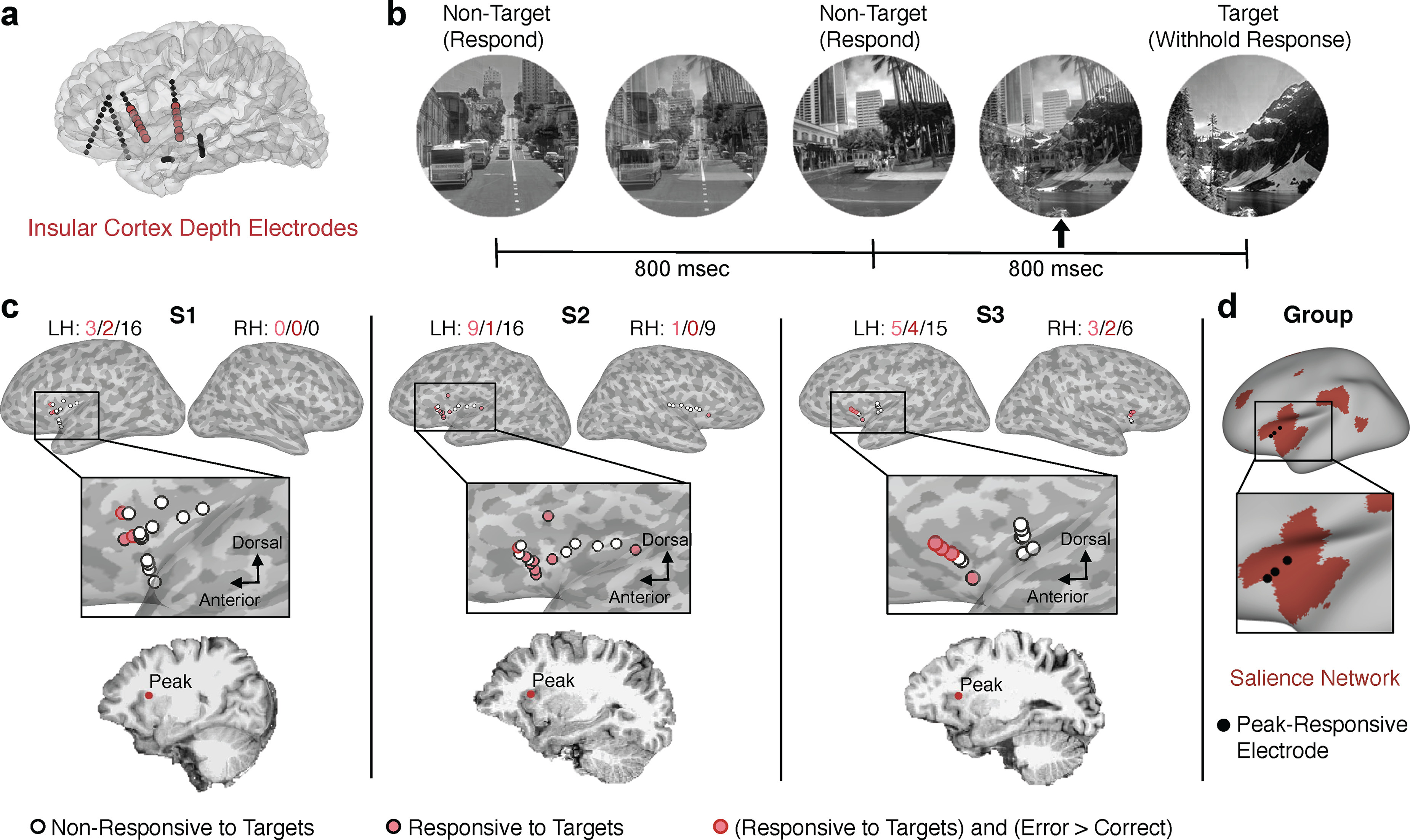
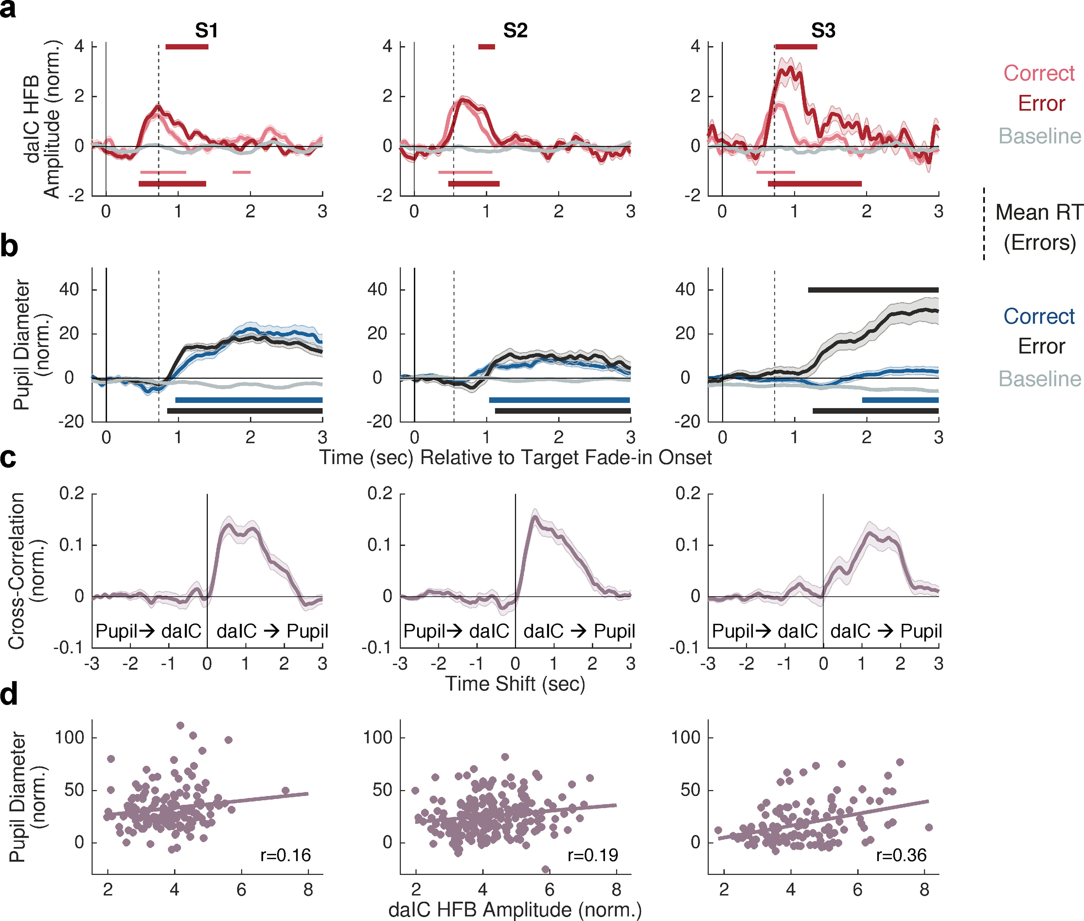
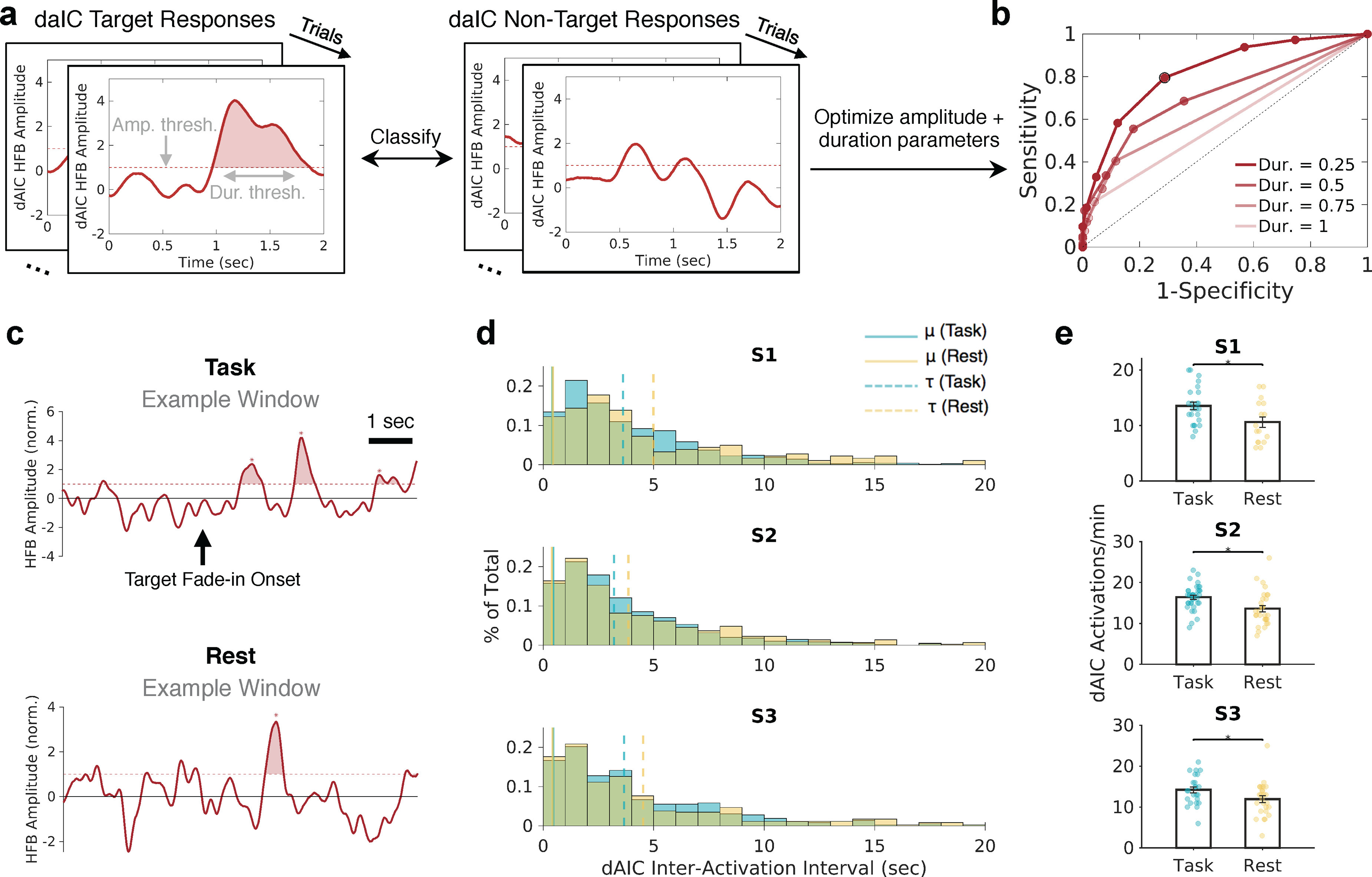
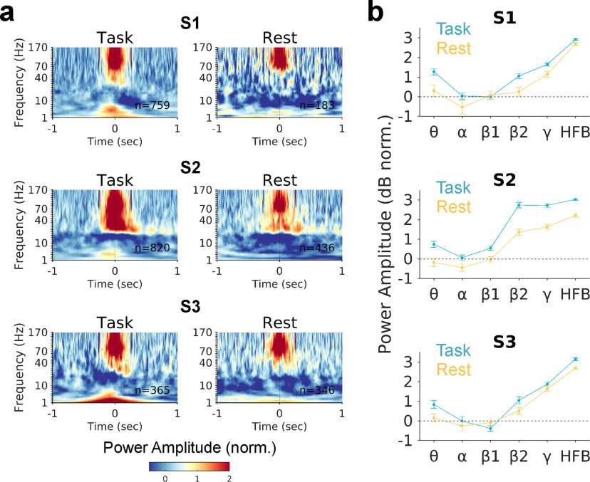
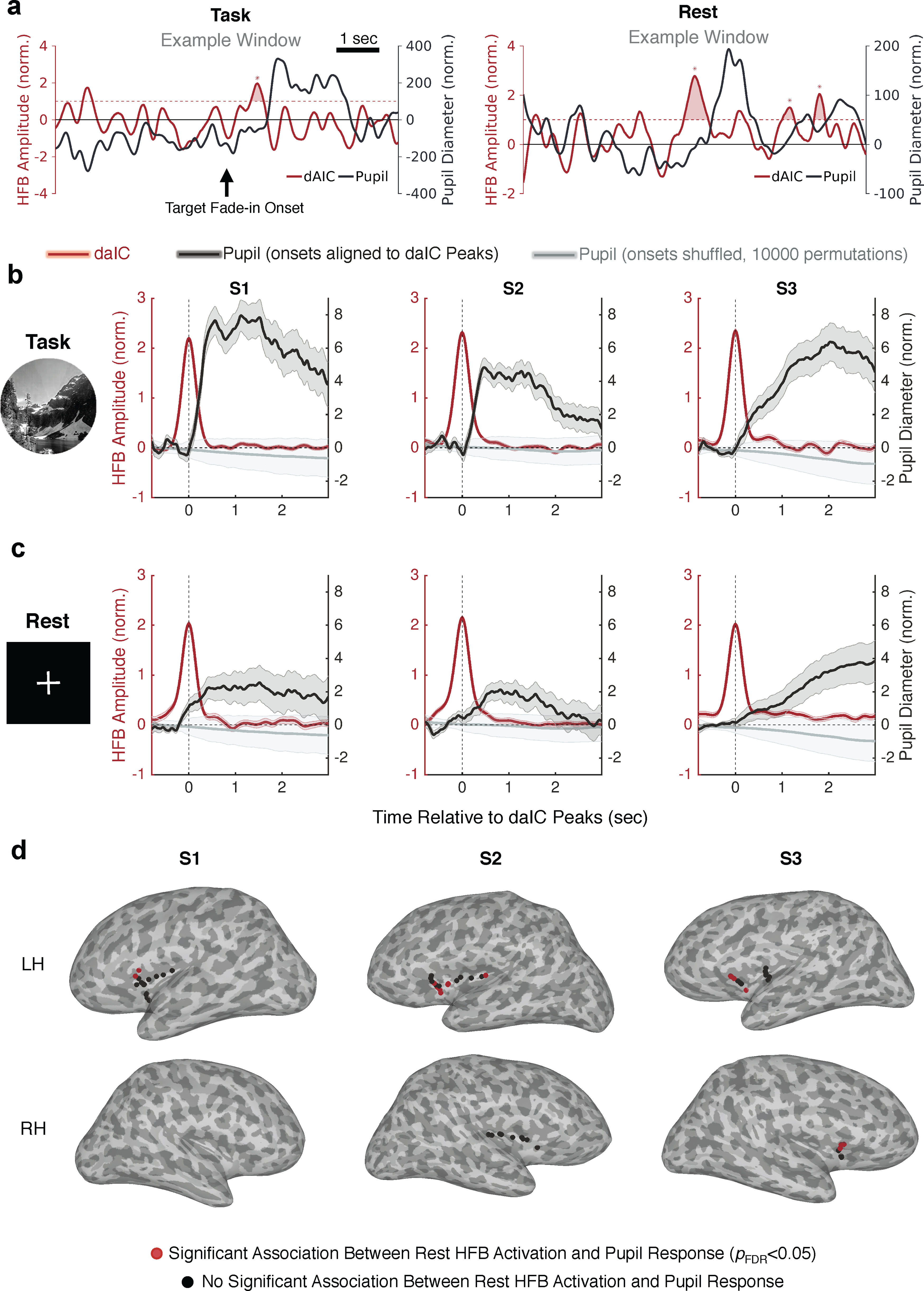
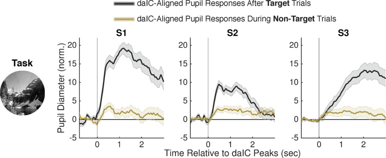
Spontaneous activations within neuronal populations can emerge similarly to "task-evoked" activations elicited during cognitive performance or sensory stimulation. We hypothesized that spontaneous activations within a given brain region have comparable functional and physiological properties to task-evoked activations. Using human intracranial EEG with concurrent pupillometry in 3 subjects (2 males, 1 female), we localized neuronal populations in the dorsal anterior insular cortex that showed task-evoked activations correlating positively with the magnitude of pupil dilation during a continuous performance task. The pupillary response peaks lagged behind insular activations by several hundreds of milliseconds. We then detected spontaneous activations, within the same neuronal populations of insular cortex, that emerged intermittently during a wakeful "resting state" and that had comparable electrophysiological properties (magnitude, duration, and spectral signature) to task-evoked activations. Critically, similar to task-evoked activations, spontaneous activations systematically preceded phasic pupil dilations with a strikingly similar temporal profile. Our findings suggest similar neurophysiological profiles between spontaneous and task-evoked activations in the human insula and support a clear link between these activations and autonomic functions measured by dynamics of pupillary dilation.SIGNIFICANCE STATEMENT Most of our knowledge about activations in the human brain is derived from studies of responses to external events and experimental conditions (i.e., "task-evoked" activations). We obtained direct neural recordings from electrodes implanted in human subjects and showed that activations emerge spontaneously and have strong similarities to task-evoked activations(e.g., magnitude, temporal profile) within the same populations of neurons. Within the dorsal anterior insula, a brain region implicated in salience processing and alertness, activations that are either spontaneous or task-evoked are coupled with brief dilations of the pupil. Our findings underscore how spontaneous brain activity, a major current focus of human neuroimaging studies aimed at developing biomarkers of disease, is relevant to ongoing physiological and possibly self-generated mental processes.

Keywords: arousal; attention; intracranial EEG; pupillometry; resting state; sympathetic.


Figures

Figure 1.
Figure 1.
Task-evoked insula and pupil responses to target stimuli in the GradCPT. a, Depth probes with electrodes implanted in the insula (red) are illustrated in an example subject. b, The GradCPT paradigm. c, Locations of insula electrodes in 3 subjects are shown on the inflated cortical surface. Electrodes were classified into those showing no significant HFB response to GradCPT target relative to nontarget stimuli (white fill, black outline), a significant HFB increased response to target relative to nontarget stimuli (pink fill), and a significant HFB increased response for target trials with correct compared with incorrect behavioral responses (red outline) (Monte Carlo p < 0.05, cluster-based permutation test corrected for number of insula electrodes within subject). The location of the electrode with strongest HFB response within each subject is shown on a 2D sagittal slice. d, Locations of peak-responsive insula electrodes within each subject displayed on the inflated fsaverage cortical surface. Red represents the salience network, based on the fMRI-based Yeo atlas of seven cortical networks.
Figure 2.
Figure 2.
Temporal coupling between task-evoked daIC activation and pupil dilation. a, Mean HFB power amplitude at peak-responsive daIC electrodes for target trials with correct behavioral responses (pink), target trials with erroneous behavioral responses (red), and nontarget trials with correct behavioral response (gray, considered as baseline). b, Mean changes in pupil diameter for correct (blue), incorrect (black), and baseline (gray) trials. a, b, Dashed vertical lines indicate the mean reaction time for target trials with erroneous behavioral responses. c, Cross-correlation (averaged across all target trials) between daIC HFB amplitude and pupil diameter. d, Trial-by-trial correlation (for all target trials) between daIC HFB amplitude and pupil diameter change. In all relevant plots, shaded error bars indicate SEM.
Figure 3.
Figure 3.
daIC activations are detected during wakeful rest but are reduced in frequency. a, Example of the presence and absence of daIC activation during GradCPT target and nontarget trials, given selected amplitude and duration thresholds. b, Example ROC curve for a single daIC electrode based on multiple pairs of candidate amplitude and duration thresholds. The pair that resulted in optimized sensitivity and specificity for trial discrimination is outlined in black. c, Example 8 s segments showing daIC activations derived from ROC curves during task and rest conditions. Shaded areas represent suprathreshold events. d, Histograms represent daIC interactivation intervals across all task and rest sessions within each subject. Based on ex-Gaussian fits, the Gaussian mean (μ, solid vertical lines) and exponential mean (τ, dotted vertical lines) are shown for task and rest. e, Activation rates in the daIC, across independent 1 min windows, for task compared with rest within each subject. *p < 0.05 (Wilcoxon rank-sum test).
Figure 4.
Figure 4.
Similar electrophysiological spectral properties between task-evoked and spontaneous daIC activation. a, Mean spectrograms, in task and wakeful resting state, aligned to HFB peaks (t = 0) defined based on ROC curves in task and rest.b, Mean power amplitude in the −50 to 50 ms window surrounding HFB peaks for the θ (4-8 Hz), α (8–12 Hz), β1(13–29 Hz), β2 (30–39 Hz), γ (40–70 Hz), and HFB ranges.
Figure 5.
Figure 5.
daIC activations precede evoked and spontaneous pupil dilations. a, Example 8 s segments showing ROC-derived daIC activations and pupil size fluctuations during task and rest conditions (examples were selected for illustrative purposes only and were not necessarily representative of mean effects). b, Pupil response (black) aligned to daIC HFB activation (red) peaks in each subject during task performance. c, Same as in a, but during wakeful rest (continuous visual fixation). Gray lines indicate the mean pupil response for 10,000 randomly shuffled distributions of daIC interactivation intervals. d, Insular cortex electrodes within each subject that either showed a significant association between HFB activation and subsequent increased pupil size (red) or did not show a significant association (black) during wakeful rest. Shaded error bars indicate SEM.
Figure 6.
Figure 6.
daIC activations precede pupil dilations during both target and nontarget trials in the GradCPT. Pupil response aligned to daIC HFB activation peaks (t = 0), split based on daIC peaks that were found after target trials (black) and during nontarget trials (olive) while patients were performing the GradCPT.

Comment in

Similar articles

Cited by

References

    1. Aron AR, Robbins TW, Poldrack RA (2014) Inhibition and the right inferior frontal cortex: one decade on. Trends Cogn Sci 18:177–185. 10.1016/j.tics.2013.12.003 - DOI - PubMed
    1. Aston-Jones G, Cohen JD (2005) An integrative theory of locus ceruleus-norepinephrine function: adaptive gain and optimal performance. Annu Rev Neurosci 28:403–450. 10.1146/annurev.neuro.28.061604.135709 - DOI - PubMed
    1. Barrett LF, Simmons WK (2015) Interoceptive predictions in the brain. Nat Rev Neurosci 16:419–429. 10.1038/nrn3950 - DOI - PMC - PubMed
    1. Bastin J, Deman P, David O, Gueguen M, Benis D, Minotti L, Hoffman D, Combrisson E, Kujala J, Perrone-Bertolotti M, Kahane P, Lachaux JP, Jerbi K (2017) Direct recordings from human anterior insula reveal its leading role within the error-monitoring network. Cereb Cortex 27:1545–1557. 10.1093/cercor/bhv352 - DOI - PubMed
    1. Beissner F, Meissner K, Bar KJ, Napadow V (2013) The autonomic brain: an activation likelihood estimation meta-analysis for central processing of autonomic function. J Neurosci 33:10503–10511. 10.1523/JNEUROSCI.1103-13.2013 - DOI - PMC - PubMed

Publication types

LinkOut - more resources