. 2020 Jul 21;117(29):17166-17176.
doi: 10.1073/pnas.2002266117. Epub 2020 Jul 6.

Impaired estrogen signaling underlies regulatory T cell loss-of-function in the chronically inflamed intestine

Affiliations

Impaired estrogen signaling underlies regulatory T cell loss-of-function in the chronically inflamed intestine

Wendy A Goodman et al. Proc Natl Acad Sci U S A. .

Abstract

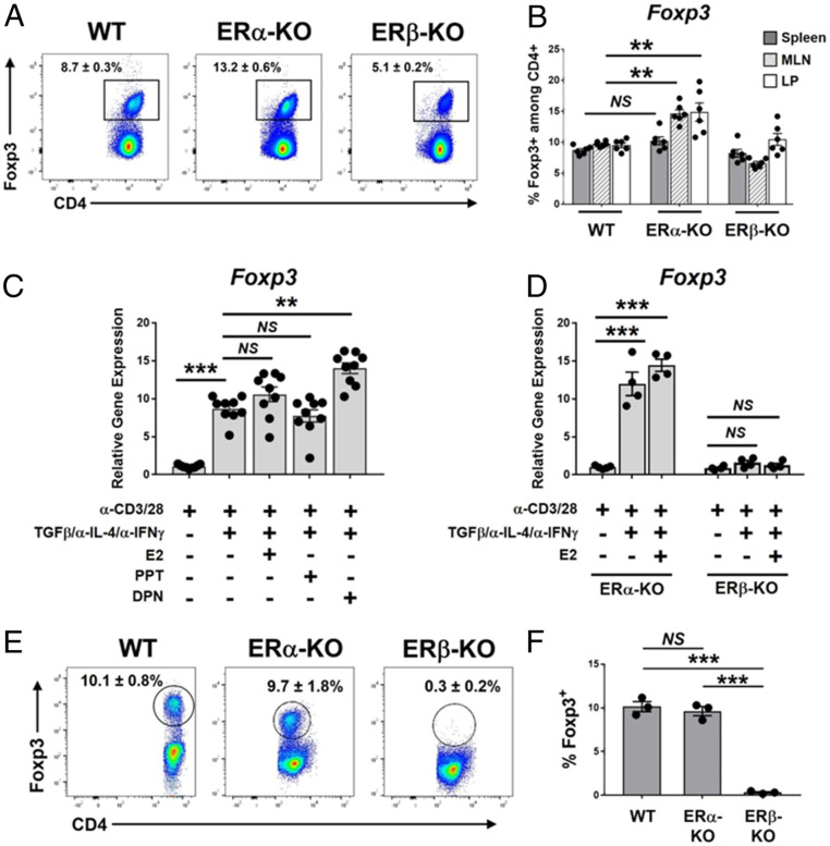
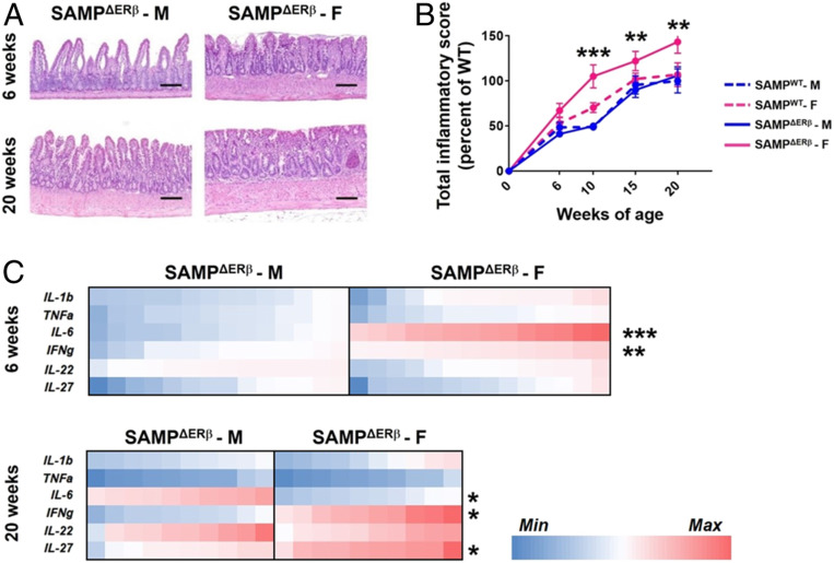
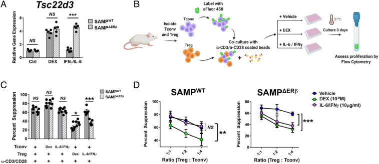
Signaling of 17β-estradiol (estrogen) through its two nuclear receptors, α and β (ERα, ERβ), is an important mechanism of transcriptional regulation. Although ERs are broadly expressed by cells of the immune system, the mechanisms by which they modulate immune responses remain poorly understood. ERβ-specific signaling is reduced in patients with chronic inflammatory diseases, including systemic lupus erythematosus and inflammatory bowel disease, and our previous work suggests that dysregulation of ERβ-specific signaling contributes to enhanced intestinal inflammation in female SAMP/YitFC mice, a spontaneous model of Crohn's disease-like ileitis. The present study builds on these prior observations to identify a nonredundant, immunoprotective role for ERβ-specific signaling in TGF-β-dependent regulatory T cell (Treg) differentiation. Using a strain of congenic SAMP mice engineered to lack global expression of ERβ, we observed dramatic, female-specific exacerbation of intestinal inflammation accompanied by significant reductions in intestinal Treg frequency and function. Impaired Treg suppression in the absence of ERβ was associated with aberrant overexpression of Tsc22d3 (GILZ), a glucocorticoid-responsive transcription factor not normally expressed in mature Tregs, and ex vivo data reveal that forced overexpression of GILZ in mature Tregs inhibits their suppressive function. Collectively, our findings identify a pathway of estrogen-mediated immune regulation in the intestine, whereby homeostatic expression of ERβ normally functions to limit Treg-specific expression of GILZ, thereby maintaining effective immune suppression. Our data suggest that transcriptional cross-talk between glucocorticoid and steroid sex hormone signaling represents an important and understudied regulatory node in chronic inflammatory disease.

Keywords: Crohn’s disease; estrogen; inflammation; inflammatory bowel disease; regulatory T cell.


Conflict of interest statement

The authors declare no competing interest.

Figures

Fig. 1.
Fig. 1.
Estrogen-mediated differentiation of peripheral Tregs requires ERβ-specific signaling. (A and B) Single-cell suspensions were prepared from ileal LP, spleens, and mLNs of 8- to 10-wk-old C57BL/6 (WT) mice or C57BL/6 mice lacking ERα or ERβ (ERα-KO, ERβ-KO; mixed male and female). Forward scatter/side scatter-gated, singlet cells were analyzed by flow cytometry for expression of CD4 and Foxp3. (A) Representative FACS histograms showing Foxp3 expression among CD4-gated lymphocytes isolated from mLNs of indicated mice. (B) Quantitation of Foxp3 expression among CD4-gated lymphocytes isolated from spleens or mLNs of indicated mice. (CF) CD4+CD44+ naïve T cells were isolated from spleens of 8- to 10-wk-old (C) C57BL/6 mice or (DF) ERα-KO or ERβ-KO mice (mixed male and female). Cells were treated ex vivo for 72 h as indicated, followed by analysis of Foxp3 expression. (C and D) Foxp3 gene expression is expressed relative to that of naïve T cells not treated with TGF-β/α-IL-4/α-IFN-γ. (E) TGF-β/α-IL-4/α-IFN-γ–treated cells were permeabilized and stained with mAbs specific to CD4 and Foxp3; representative flow cytometry histograms are shown. (F) Quantitation of results shown in E, representing the percentage of CD4+ cells expressing Foxp3. Data represent the mean ± SEM (NS, not significant; **P ≤ 0.01; ***P ≤ 0.001, n = 3 to 9 per group).
Fig. 2.
Fig. 2.
Female CD patients exhibit reduced ERβ-specific signaling. Gene expression of ERα and ERβ was analyzed in (A) ileal tissue biopsy samples or (B) peripheral blood T cells from indicated donors by qPCR. (A) Relative gene expression of ERα or ERβ is expressed compared to Ctrl males. (B) Ratio of ERα:ERβ expression is expressed for Tconv or Treg cells. Each circle represents an individual donor. Data represent the mean ± SEM (NS, not significant; **P ≤ 0.01; ***P ≤ 0.001, n = 5 to 11 per group).
Fig. 3.
Fig. 3.
Global deletion of ERβ exacerbates experimental ileitis in female SAMP mice. SAMP mice lacking global expression of ERβ (SAMPΔERβ mice) were generated by back-crossing SAMP mice with ERβ-KO mice. (A) Representative H&E-stained proximal ileal tissue from 6- and 20-wk-old SAMPΔERβ mice. (Scale bars, 10 μM.) (B) Total inflammatory scores are shown for SAMPΔERβ mice at 6, 10, 15, and 20 wk of age. Data represent the mean ± SEM (**P ≤ 0.01; ***P ≤ 0.001, n = 8 to 14 per group). (C) Relative gene expression of selected proinflammatory genes in full-thickness proximal ileal tissues isolated from 6- or 20-wk old SAMPΔERβ mice is expressed as a heat map. F, female; M, male. *P ≤ 0.05.
Fig. 4.
Fig. 4.
Female SAMPΔERβ mice show impaired Treg differentiation and function. (A) Representative FACS histogram showing percentages of CD4+-gated, Foxp3-expressing T cells isolated from mLNs of indicated mice. (B) Quantitation of results shown in A. (C) Relative expression of Treg-associated genes in full-thickness proximal ileal tissues isolated from indicated mice (all 12 to 15 wk of age). (D) Representative FACS plots showing e450 dye dilution of proliferating CD4+CD25 Tconv cells cultured alone (Upper) or cocultured (Lower) with CD4+CD25highCD127lo-expressing Tregs (4:1 Tconv:Treg) isolated from mLNs of male or female SAMP-ERβ-KO mice. (E) Quantitation of results shown in D. (F) Percentage of CD39+/CD73+ coexpressing Tregs from indicated mice, assessed by FACS. Data represents the mean ± SEM (NS, not significant; *P ≤ 0.05; **P ≤ 0.01; ***P ≤ 0.001, n = 3 to 8 per group).
Fig. 5.
Fig. 5.
Impaired expression of ERβ allows aberrant overexpression of Tsc22d3 (GILZ) in Tregs. (A and B) CD4+CD25highCD127lo Tregs were isolated from mLNs of SAMPΔERβ mice and SAMPWT littermate controls by FACS sorting. (A) Tregs were analyzed by next-generation RNA-seq for changes to the global transcriptome, and a dendrogram shows the top 50-most differentially expressed genes by interaction-contrast analysis. (B) Gene expression of Tsc22d3 was analyzed among mLN-isolated Tregs from indicated mice by qPCR. (C and D) CD4+CD44+ naïve T cells were isolated from spleens of SAMPΔERβ male (C) or female (D) mice, then cultured ex vivo with α-CD3/CD28, TGF-β, and neutralizing antibodies α-IL-4 and α-IFN-γ for 5 d. Data represents relative gene expression of Tsc22d3, compared to day 0, in cells from indicated mice. (E) Gene expression of TSC22D3 was analyzed among peripheral blood-derived CD4+CD25highCD127lo Tregs isolated from Ctrl or CD patients. Data represents the mean ± SEM (NS, not significant; **P ≤ 0.01; ***P ≤ 0.001, n = 3 to 8 per group). (F and G) Amnis Imagestream cytometry was used to visualize colocalize ERβ and GILZ in peripheral blood-derived CD4+CD25highCD127lo Tregs isolated from Ctrl or CD patients. (F) Representative Imagestream data from indicated patients is shown; similarity score indicates correlation between MFI (ERβ) and MFI (GILZ). (G) Correlation analysis showing ΔMFI (GILZ) versus ΔMFI (ERβ) for Tregs isolated from male versus female CD patients.
Fig. 6.
Fig. 6.
Treg-specific expression of GILZ impairs suppressive function. (A) CD4+CD25highCD127lo Tregs were isolated from mLNs of SAMPΔERβ or SAMPWT mice (mixed male/female) and treated ex vivo as indicated (48 h). Gene expression of Tsc22d3 was determined by qPCR and is expressed relative to Ctrl (untreated) SAMPWT expression values. (BD) CD4+CD25 Tconv were isolated from spleens of SAMPWT mice (mixed male/female) and cocultured ex vivo with CD4+CD25highCD127lo Tregs isolated from mLNs of SAMPWT or SAMPΔERβ mice as indicated. Either DEX or IL-6/IFN-γ was added to cocultures on day 0. (B) Schematic showing experimental design for Treg suppression assays ± Dex or IL-6/IFN-γ. Figure was created using BioRender. (C) Percent Treg suppression of Tconv proliferation is expressed for the indicated conditions (1:1 Treg:Tconv ratio). (D) Percent Treg suppression of Tconv proliferation is expressed for indicated ratios of Treg:Tconv. Data represent the mean ± SEM (NS, not significant; *P ≤ 0.05; **P ≤ 0.01; ***P ≤ 0.001, n = 3 to 5 per group).
Fig. 7.
Fig. 7.
Graphical summary of proposed ERα-ERβ-GILZ signaling axis in Tregs. (Left) In the healthy, noninflamed intestinal mucosae, ERα and ERβ are expressed at physiological levels by immune cells including Tregs. Soluble E2 diffuses across the plasma membrane and binds ERα and ERβ in the cytoplasm, after which ERα/ERβ heterodimers form. These dimers then translocate to the nucleus and bind estrogen response elements (EREs) in target gene promoters. ERβ-containing dimers inhibit the transcriptional activation of Tsc22d3 (GILZ) in Tregs under noninflamed conditions. (Right) In the chronically inflamed intestine, expression of ERβ is significantly reduced in Tregs. Additional signals from cytokines such as IL-6 and IFN-γ contribute to a chronic inflammatory environment and may interact with ER signaling to influence target gene transcription. In the absence of ERβ, ERα/ERα homodimers permit Tsc22d3 transcription in Tregs, leading to loss of functional Treg suppression. Figure was created using BioRender.

Similar articles

Cited by

References

    1. Fish E. N., The X-files in immunity: Sex-based differences predispose immune responses. Nat. Rev. Immunol. 8, 737–744 (2008). - PMC - PubMed
    1. Klein S. L., Flanagan K. L., Sex differences in immune responses. Nat. Rev. Immunol. 16, 626–638 (2016). - PubMed
    1. Ortizo R. et al. ., Exposure to oral contraceptives increases the risk for development of inflammatory bowel disease: A meta-analysis of case-controlled and cohort studies. Eur. J. Gastroenterol. Hepatol. 29, 1064–1070 (2017). - PubMed
    1. Khalili H. et al. ., Association between long-term oral contraceptive use and risk of Crohn’s disease complications in a nationwide study. Gastroenterology 150, 1561–1567.e1 (2016). - PMC - PubMed
    1. Khalili H. et al. ., Oral contraceptives, reproductive factors and risk of inflammatory bowel disease. Gut 62, 1153–1159 (2013). - PMC - PubMed

Publication types

MeSH terms