. 2020 Jul 21;117(29):17240-17248.
doi: 10.1073/pnas.1914430117. Epub 2020 Jul 6.

A molecular mechanism for probabilistic bet hedging and its role in viral latency

Affiliations

A molecular mechanism for probabilistic bet hedging and its role in viral latency

Sonali Chaturvedi et al. Proc Natl Acad Sci U S A. .

Abstract

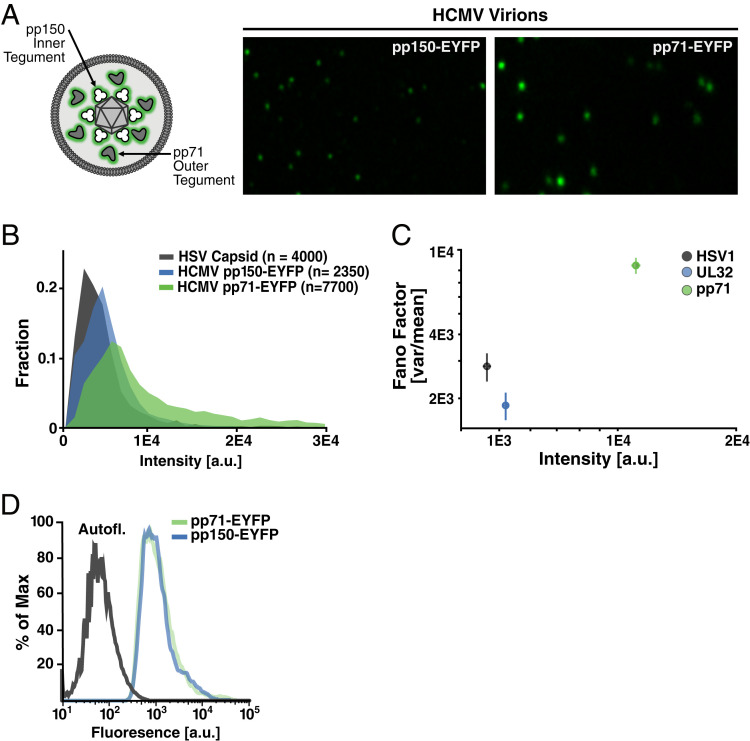
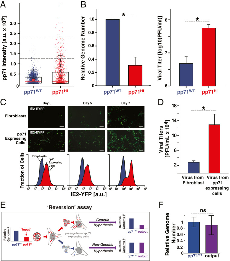
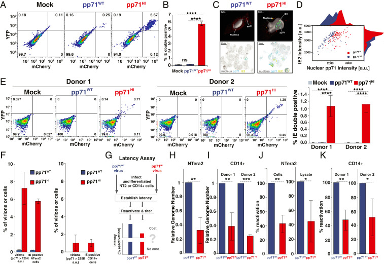
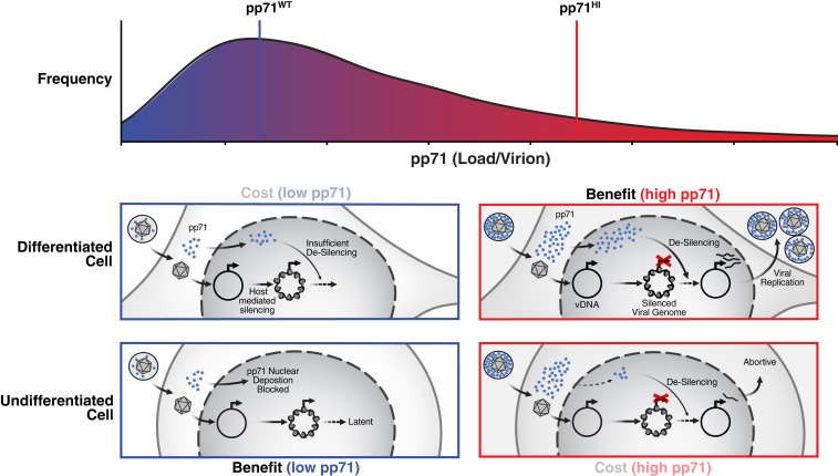
Probabilistic bet hedging, a strategy to maximize fitness in unpredictable environments by matching phenotypic variability to environmental variability, is theorized to account for the evolution of various fate-specification decisions, including viral latency. However, the molecular mechanisms underlying bet hedging remain unclear. Here, we report that large variability in protein abundance within individual herpesvirus virion particles enables probabilistic bet hedging between viral replication and latency. Superresolution imaging of individual virions of the human herpesvirus cytomegalovirus (CMV) showed that virion-to-virion levels of pp71 tegument protein-the major viral transactivator protein-exhibit extreme variability. This super-Poissonian tegument variability promoted alternate replicative strategies: high virion pp71 levels enhance viral replicative fitness but, strikingly, impede silencing, whereas low virion pp71 levels reduce fitness but promote silencing. Overall, the results indicate that stochastic tegument packaging provides a mechanism enabling probabilistic bet hedging between viral replication and latency.

Keywords: fate selection; herpesvirus; latency; stochastic variability; tegument.


Conflict of interest statement

The authors declare no competing interest.

Figures

Fig. 1.
Fig. 1.
Substantial virion-to-virion heterogeneity in the level of the HCMV major tegument transactivator protein pp71. (A, Left) Schematic of HCMV virion showing relative locations of pp150 (UL32), which is capsid-associated, in the inner tegument, and pp71 (UL82), which is capsid-unassociated, in the outer tegument. (A, Right) Representative superresolution fluorescence micrographs of purified, infectious, recombinant HCMV virion particles where tegument proteins are genetically fused to EYFP: pp150-EYFP (Left), pp71-EYFP (Right) (pixel size; 40 nm). (B) Quantification of pp71 and pp150 abundance in individual virion particles relative to the HSV-1 VP26-GFP “molecular ruler” (900 copies per virion). (C) Normalized variance (Fano factor; σ2/μ) versus mean abundance (<intensity>) in pp71, pp150, and HSV-1 capsid in the virion populations. Error bars were estimated by bootstrapping the data with n = 1,000 particles per sample, 150 times. (D) Intracellular levels of tegument factors EYFP-pp71 and pp150-EYFP in infected human fibroblasts quantified by flow cytometry (Left).
Fig. 2.
Fig. 2.
The “advantage” of high pp71: high virion pp71 levels enhance HCMV replicative fitness. (A) Box-whisker plot of pp71 levels (by EYFP intensity) in purified virion particles quantified by superresolution microscopy: pp71WT EYFP virion particles (purple, n = 5,519); pp71HI EYFP virion particles (red, n = 2,463). Analysis was restricted to particles 100–300 nm in diameter. The red square depicts the mean μ, red line the median, the box encloses values between the first and third quantiles (25–75%), and the whiskers (error bars) show the minimum and maximum intensities. The horizontal-dashed lines represent intensity thresholds of 1.25 × 10⁵ a.u. (dark gray) and 2.25 × 10⁵ a.u. (light gray). For pp71WT, 0.25% of virions >1.25 × 10⁵ a.u. and 0% of virions >2.25 × 10⁵ a.u.; for pp71HI, 7.1% of virions >1.25 × 10⁵ a.u. and 1% of virions >2.25 × 10⁵ a.u. (these percentage are shown in Fig. 3F). (B, Left) qRT-PCR analysis of viral genome copy number in pp71HI and pp71WT stocks after stocks were normalized to have equivalent infectivity by infectious units (*P < 0.05, two-tailed t test). (B, Right) Infectivity of same pp71HI and pp71WT stocks, as measured by TCID50, after stocks were matched to have equivalent genome copy numbers (*P < 0.05, two-tailed t test). (C, Upper) Confocal micrographs of IE2-YFP reporter-virus infections (MOI 0.01) on human fibroblasts (HFFs) or a pp71-expressing HFF line (WF28). (Scale bars, 200 µm.) (C, Lower) Flow cytometry analysis of lytic IE2-YFP expression on indicated days. (D) Viral replication (titer) quantified by TCID50 7 d.p.i. of HFFs or pp71-expressing HFFs. (E) Schematic of “Reversion assay” to test if pp71HI virus harbors secondary mutations that influence its phenotype. If pp71HI phenotype has a genetic component, lower genome-copy number (i.e., higher infectious particle-to-genome ratio) relative to pp71WT will be retained and selected for due to its replicative advantage. In contrast, if the pp71HI phenotype is nongenetic, excess packaged pp71 will not be retained on low passage in naïve cells and phenotype will revert to pp71WT (i.e., genome-copy number and infectious particle-to-genome ratio equal to pp71WT). (F) qPCR analysis of virus output from the “reversion assay” (i.e., pp71HI virus after low passage in HFF). Titer of the resulting virus was measured by TCID50, matched to WT (as in Fig. 2B), and viral genomes in the MOI-matched isolate then quantified by qPCR (difference is not significant by t test). ns, not significant. (Magnification: 20× obj.)
Fig. 3.
Fig. 3.
The “cost” of high pp71: high virion pp71 levels overcome nuclear exclusion in undifferentiated cells and impede establishment of viral silencing. (A) Flow cytometry analysis of undifferentiated NTera2 cells infected with dual-reporter TB40E-IE-mCherry-EYFP. Cells were either mock infected (Left), infected with pp71WT virus (Center), or with pp71HI virus (Right) at MOI = 3. (B) Quantification of % IE double-positive population for three biological replicates from the flow cytometry data shown in Fig. 3A (P values from Student’s t test: ****<0.0001). (C) Representative confocal immunofluorescence micrographs of infected NTera2 cells (MOI = 3) assayed 6 h.p.i. with either pp71WT or pp71HI virus expressing IE2-YFP. Two-dimensional images along with a single confocal plane from a three-dimensional (3D) z-stack are shown along with a 3D reconstruction of the cell nucleus (solid). pp71 (stained via α-pp71 antibody) is visible as fluorescent puncta (teal) proximal to IE2-YFP puncta (green). Extranuclear fluorescence (red) is due to cytoplasmic autofluorescence. (D) Image-based quantification of pp71 and IE2 intensity levels in cells exhibiting both IE2 expression and nuclear pp71 levels for cells infected with pp71WT (purple; 105 cells) or pp71HI (red; 109 cells). Histograms on axes are derived from projecting the dot-plot data onto respective axis. (E, Left) Flow cytometry analysis of donor-derived human CD14+ primary monocytes infected with dual-reporter TB40E-IE-mCherry-EYFP (MOI = 2), 6 h.p.i., for two donors. Cells were either mock infected, infected with pp71WT virus or pp71HI virus. (E, Right) Quantification of %IE double-positive CD14(+) monocytes per donor, as assayed by flow cytometry 6 h.p.i. (P values as in B). (F) Comparative analysis of percentage of virions with high pp71 levels (from Fig. 2A) vs. cells with IE expression (Fig. 3 C and E). (G) Schematic of the latency assay for pp71WT and pp71HI virus. Briefly, undifferentiated NTera2 or CD14+ cells are infected with either pp71WT or pp71HI virus, latency established over 4–10 d, and virus then reactivated from latency. Latency is quantified by qPCR for viral genomes as well as titering on HFF after reactivation. (H) qPCR quantification of latent CMV genomes 4 d after TB40E infection of NTera2 cells. (I) qPCR quantification of latent CMV genomes 10 d after TB40E infection of CD14+ monocytes. (J) Analysis of latent reactivation in NTera2 cells (initial infection with pp71WT or pp71HI TB40E-IE-mCherry-YFP virus at MOI = 3). Four days postinfection, NTera2 cells were treated with TSA for 24 h, washed three times, and serially diluted and cocultured with HFFs for 10 d and plaque-forming units/mL then calculated by TCID50. NTera2 cell lysate was titered in parallel for 10 d. Average of three biological replicates shown with pp71WT titers normalized to 100% reactivation. (K) Analysis of latent reactivation in human primary CD14+ monocytes (initial infection with pp71WT or pp71HI TB40E-IE-mCherry-YFP virus at MOI = 2). Ten days postinfection, CD14+ cells were cocultured with HFFs (10-fold serial dilution) supplemented with reactivation media (IL3, IL6, G-CSF, GM-CSF) for 15 d and viral titers then analyzed by TCID50. The experiment was performed for two donors with three biological replicates and shown with pp71WT titers normalized to 100% reactivation. (P value <0.05 was considered statistically significant: *<0.05, **<0.01, ***<0.001, ****<0.0001, two-tailed t test).
Fig. 4.
Fig. 4.
Summary model: super-Poissonian variability in tegument abundance enables bet hedging between replication and latency. In differentiated cells (Middle), pp71HI virions (Right) have an “advantage” over virions with low pp71: pp71HI enables desilencing and enhanced replicative fitness compared to pp71WT virions that are more likely to generate a silenced infection. In undifferentiated cells (Bottom), pp71HI virions (Right) have a “cost” as they impede viral silencing and reduce establishment of latency. In undifferentiated cells, virions with low pp71 levels promote establishment of viral silencing and latency.

Similar articles

Cited by

References

    1. Cohen D., Optimizing reproduction in a randomly varying environment. J. Theor. Biol. 12, 119–129 (1966). - PubMed
    1. Balaban N. Q., Persistence: Mechanisms for triggering and enhancing phenotypic variability. Curr. Opin. Genet. Dev. 21, 768–775 (2011). - PubMed
    1. Golding I., Infection by bacteriophage lambda: An evolving paradigm for cellular individuality. Curr. Opin. Microbiol. 43, 9–13 (2018). - PMC - PubMed
    1. Symmons O., Raj A., What’s luck got to do with it: Single cells, multiple fates, and biological nondeterminism. Mol. Cell 62, 788–802 (2016). - PMC - PubMed
    1. Balaban N. Q., Merrin J., Chait R., Kowalik L., Leibler S., Bacterial persistence as a phenotypic switch. Science 305, 1622–1625 (2004). - PubMed

Publication types

Substances